Skip directly to site content Skip directly to page options Skip directly to A-Z link Skip directly to A-Z link Skip directly to A-Z link

Disclaimer: Early release articles are not considered as final versions. Any changes will be reflected in the online version in the month the article is officially released.

Volume 31, Number 11—November 2025

Research Letter

Mortality Event in Rainbow Snakes Linked to Snake Fungal Disease, United States

Dane A. ConleyComments to Author , Gaëlle Blanvillain, Jaimie L. Miller, Kate E. Langwig, John D. Kleopfer, Jeffrey M. Lorch1, and Joseph R. Hoyt1
Author affiliation: Virginia Polytechnic Institute and State University, Blacksburg, Virginia, USA (D.A. Conley, G. Blanvillain, K.E. Langwig, J.R. Hoyt); University of Wisconsin–Madison, School of Veterinary Medicine, Madison, Wisconsin, USA (J.L. Miller); Virginia Department of Wildlife Resources, Henrico, Virginia, USA (J.D. Kleopfer); US Geological Survey, National Wildlife Health Center, Madison, Wisconsin, USA (J.M. Lorch)

Main Article

Figure 1

Encounters with dead and live snakes over time from a study of a mortality event in rainbow snakes linked to SFD, United States. A) Map of locations where dead Farancia erytrogramma rainbow snakes were observed during 2013–2023. Exact locations were jittered to obscure sensitive habitat. B) Stacked bar plot showing dead F. erytrogramma snakes found per kilometer in the Back Bay region of Virginia and North Carolina. Dead snakes characterized based on strength of evidence that they died from SFD: suspected, snakes with characteristic SFD lesions in photographs but no screening for Ophidiomyces ophidiicola performed; presumptive, snakes with characteristic SFD lesions, detection of O. ophidiicola, but no necropsies performed; confirmed, snakes with characteristic SFD lesions, detection of O. ophidiicola, and characteristic histologic lesions confirmed through histology; O. ophidiicola detected, snakes with no apparent lesions but tested positive for O. ophidiicola; no lesions, snakes with no apparent lesions in photographs and not tested for O. ophidiicola; unknown, snakes with no photographs of the dorsal and ventral sides. C) Number of live F. erytrogramma snakes encountered in the field per kilometer of transect surveyed with an incorporated 20-coverboard array during 2020–2023 (n = 19; zero-inflated Poisson log-scale year coefficient = −0.482 ± 0.226; p = 0.033) (Appendix Table 3). Total kilometers surveyed per year represented above survey data points. Color shading corresponds to mean infection severity (red is more severe than orange) during sampling event when the species was detected. Gray indicates sampling events without live rainbow snakes detected. SFD, snake fungal disease.

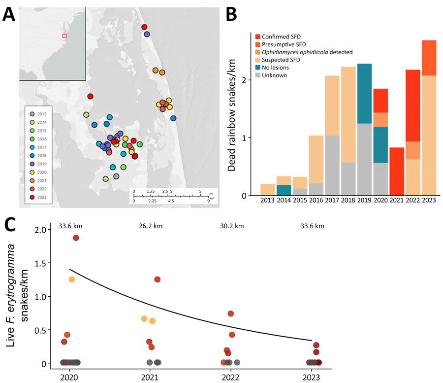
Figure 1. Encounters with dead and live snakes over time from a study of a mortality event in rainbow snakes linked to SFD, United States. A) Map of locations where dead Farancia erytrogramma rainbow snakes were observed during 20132023. Exact locations were jittered to obscure sensitive habitat. B) Stacked bar plot showing dead F. erytrogramma snakes found per kilometer in the Back Bay region of Virginia and North Carolina. Dead snakes characterized based on strength of evidence that they died from SFD: suspected, snakes with characteristic SFD lesions in photographs but no screening for Ophidiomyces ophidiicola performed; presumptive, snakes with characteristic SFD lesions, detection of O. ophidiicola, but no necropsies performed; confirmed, snakes with characteristic SFD lesions, detection of O. ophidiicola, and characteristic histologic lesions confirmed through histology; O. ophidiicola detected, snakes with no apparent lesions but tested positive for O. ophidiicola; no lesions, snakes with no apparent lesions in photographs and not tested for O. ophidiicola; unknown, snakes with no photographs of the dorsal and ventral sides. C) Number of live F. erytrogramma snakes encountered in the field per kilometer of transect surveyed with an incorporated 20-coverboard array during 2020–2023 (n = 19; zero-inflated Poisson log-scale year coefficient = −0.482 ± 0.226; p = 0.033) (Appendix Table 3). Total kilometers surveyed per year represented above survey data points. Color shading corresponds to mean infection severity (red is more severe than orange) during sampling event when the species was detected. Gray indicates sampling events without live rainbow snakes detected. SFD, snake fungal disease.

Main Article

1These authors contributed equally to this article.

Page created: October 01, 2025
Page updated: December 04, 2025
Page reviewed: December 04, 2025
The conclusions, findings, and opinions expressed by authors contributing to this journal do not necessarily reflect the official position of the U.S. Department of Health and Human Services, the Public Health Service, the Centers for Disease Control and Prevention, or the authors' affiliated institutions. Use of trade names is for identification only and does not imply endorsement by any of the groups named above.
file_external