Skip directly to site content Skip directly to page options Skip directly to A-Z link Skip directly to A-Z link Skip directly to A-Z link

Disclaimer: Early release articles are not considered as final versions. Any changes will be reflected in the online version in the month the article is officially released.

Volume 31, Number 11—November 2025

Research Letter

Mortality Event in Rainbow Snakes Linked to Snake Fungal Disease, United States

Dane A. ConleyComments to Author , Gaëlle Blanvillain, Jaimie L. Miller, Kate E. Langwig, John D. Kleopfer, Jeffrey M. Lorch1, and Joseph R. Hoyt1
Author affiliation: Virginia Polytechnic Institute and State University, Blacksburg, Virginia, USA (D.A. Conley, G. Blanvillain, K.E. Langwig, J.R. Hoyt); University of Wisconsin–Madison, School of Veterinary Medicine, Madison, Wisconsin, USA (J.L. Miller); Virginia Department of Wildlife Resources, Henrico, Virginia, USA (J.D. Kleopfer); US Geological Survey, National Wildlife Health Center, Madison, Wisconsin, USA (J.M. Lorch)

Main Article

Figure 2

Variation in Ophidiomyces ophidiicola infection among snake species from a study of a mortality event in rainbow snakes linked to snake fungal disease, United States. Sampling results during spring (January−June) in 2020–2023 in the Back Bay watershed in Virginia and North Carolina. Black circles and error bars indicate mean lesion severity with 95% CIs. A) Each colored point represents an individual snake sampled and whether it was positive (1) or negative (0) for O. ophidiicola. Data points are slightly jittered for visualization purposes. B) Summed lesion severity values accounting for lesion size, lesion progression, and proportion of snake affected (Appendix). Snakes without lesions were omitted.

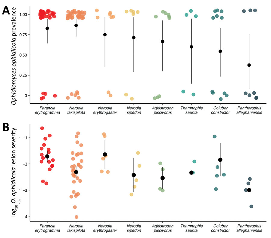
Figure 2. Variation in Ophidiomyces ophidiicola infection among snake species from a study of a mortality event in rainbow snakes linked to snake fungal disease, United States. Sampling results during spring (January−June) in 20202023 in the Back Bay watershed in Virginia and North Carolina. Black circles and error bars indicate mean lesion severity with 95% CIs. A) Each colored point represents an individual snake sampled and whether it was positive (1) or negative (0) for O. ophidiicola. Data points are slightly jittered for visualization purposes. B) Summed lesion severity values accounting for lesion size, lesion progression, and proportion of snake affected (Appendix). Snakes without lesions were omitted.

Main Article

1These authors contributed equally to this article.

Page created: October 01, 2025
Page updated: December 04, 2025
Page reviewed: December 04, 2025
The conclusions, findings, and opinions expressed by authors contributing to this journal do not necessarily reflect the official position of the U.S. Department of Health and Human Services, the Public Health Service, the Centers for Disease Control and Prevention, or the authors' affiliated institutions. Use of trade names is for identification only and does not imply endorsement by any of the groups named above.
file_external