Volume 31, Number 11—November 2025
Dispatch
Bjerkandera spp. Pulmonary Infection in Immunocompromised Hosts, Germany
Rosanne Sprute , Danila Seidel, Katrin Mehler, Zoé Westhues, Sarina K. Butzer, Jannik Stemler, Oliver A. Cornely, and Philipp Koehler
Figure 3
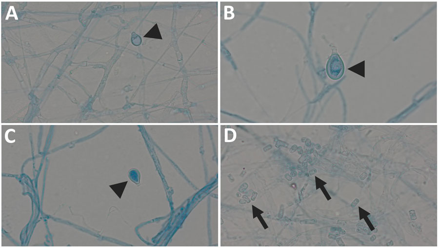
Figure 3. Images of Bjerkandera spp. formations from study of Bjerkandera spp. pulmonary infection in immunocompromised hosts, Germany. Slides are of lactophenol preparation (original magnification ×1,000). Bjerkandera spp. form white, yellowish-white, or tan colonies with a cottony to woolly texture on malt extract agar. The hyphae can be branched. Thin-walled, rectangular arthroconidia are formed via schizolytic dehiscence. In addition, ellipsoidal chlamydospores <10 µm long may develop. Arrowheads indicate chlamydospores (A–C), arrows indicate arthroconidia (D).
Page created: September 26, 2025
Page updated: December 10, 2025
Page reviewed: December 10, 2025
The conclusions, findings, and opinions expressed by authors contributing to this journal do not necessarily reflect the official position of the U.S. Department of Health and Human Services, the Public Health Service, the Centers for Disease Control and Prevention, or the authors' affiliated institutions. Use of trade names is for identification only and does not imply endorsement by any of the groups named above.