Volume 31, Number 11—November 2025
Dispatch
Bjerkandera spp. Pulmonary Infection in Immunocompromised Hosts, Germany
Rosanne Sprute , Danila Seidel, Katrin Mehler, Zoé Westhues, Sarina K. Butzer, Jannik Stemler, Oliver A. Cornely, and Philipp Koehler
Figure 2
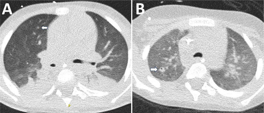
Figure 2. Chest computed tomography scan of a 4-year-old male patient with acute myeloid leukemia in study of Bjerkandera spp. pulmonary infection in immunocompromised hosts, Germany. The patient experienced fever unresponsive to antimicrobial treatment. A) Imaging revealed nodular infiltrates and surrounding ground-glass opacities in both lungs (arrow). Bjerkandera spp. was identified from tracheal aspiration. B) Follow-up computed tomography scan after 4 weeks demonstrated regressive nodular lesions and the formation of a cavity in the right upper lobe (arrow).
Page created: September 26, 2025
Page updated: December 10, 2025
Page reviewed: December 10, 2025
The conclusions, findings, and opinions expressed by authors contributing to this journal do not necessarily reflect the official position of the U.S. Department of Health and Human Services, the Public Health Service, the Centers for Disease Control and Prevention, or the authors' affiliated institutions. Use of trade names is for identification only and does not imply endorsement by any of the groups named above.