Skip directly to site content Skip directly to page options Skip directly to A-Z link Skip directly to A-Z link Skip directly to A-Z link

Disclaimer: Early release articles are not considered as final versions. Any changes will be reflected in the online version in the month the article is officially released.

Volume 31, Number 7—July 2025

Dispatch

Multisystemic Disease and Septicemia Caused by Presumptive Burkholderia pseudomallei in American Quarter Horse, Florida, USA

Jason J. ThorntonComments to Author , John F. Roberts, David P. AuCoin, and Apichai Tuanyok
Author affiliation: University of Florida College of Veterinary Medicine, Gainesville, Florida, USA (J.J. Thornton, J.F. Roberts, A. Tuanyok); University of Nevada School of Medicine, Reno, Nevada, USA (D.P. AuCoin)

Main Article

Figure 2

Photomicrography of tissues from study of multisystemic disease and septicemia caused by presumptive Burkholderia pseudomallei in American quarter horse, Florida, USA. Photomicrographs show hematoxylin and eosin staining of submandibular lymph node (LN) (A, C, E) and lung (B, D) and Gram stain of the submandibular LN (F). A) Submandibular LN shows multifocal to coalescing pyogranulomas. Original magnification ×2. B) The lung contains a parabronchial abscess (star). Original magnification ×2. C) The submandibular LN pyogranuloma contains necrotic debris, suppurative inflammation, and numerous hemosiderin laden macrophages, multinucleated giant cells surrounded by fibrosis. Original magnification ×20. D) Macrophages and neutrophils in the pulmonary abscess contain a mixed population of 1–2 μm coccobacilli. Original magnification ×40. E) The submandibular node neutrophils and macrophages contain numerous intracellular 1–2 μm bacilli (red arrows). Original magnification ×40. F) The bacilli in the submandibular lymph node are diffusely gram-negative (black arrows). Original magnification ×30.

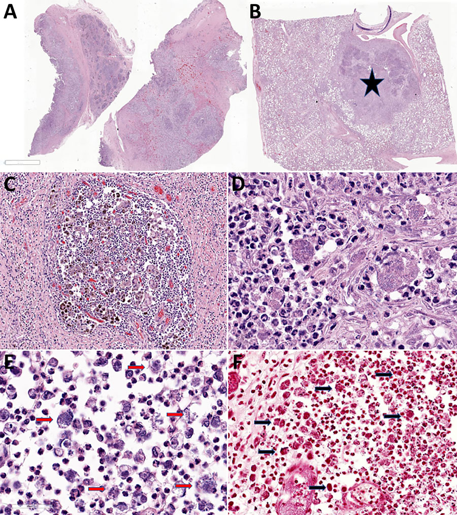
Figure 2. Photomicrography of tissues from study of multisystemic disease and septicemia caused by presumptive Burkholderia pseudomallei in American quarter horse, Florida, USA. Photomicrographs show hematoxylin and eosin staining of submandibular lymph node (LN) (A, C, E) and lung (B, D) and Gram stain of the submandibular LN (F). A) Submandibular LN shows multifocal to coalescing pyogranulomas. Original magnification ×2. B) The lung contains a parabronchial abscess (star). Original magnification ×2. C) The submandibular LN pyogranuloma contains necrotic debris, suppurative inflammation, and numerous hemosiderin laden macrophages, multinucleated giant cells surrounded by fibrosis. Original magnification ×20. D) Macrophages and neutrophils in the pulmonary abscess contain a mixed population of 1–2 μm coccobacilli. Original magnification ×40. E) The submandibular node neutrophils and macrophages contain numerous intracellular 1–2 μm bacilli (red arrows). Original magnification ×40. F) The bacilli in the submandibular lymph node are diffusely gram-negative (black arrows). Original magnification ×30.

Main Article

Page created: May 01, 2025
Page updated: June 17, 2025
Page reviewed: June 17, 2025
The conclusions, findings, and opinions expressed by authors contributing to this journal do not necessarily reflect the official position of the U.S. Department of Health and Human Services, the Public Health Service, the Centers for Disease Control and Prevention, or the authors' affiliated institutions. Use of trade names is for identification only and does not imply endorsement by any of the groups named above.
file_external