Skip directly to site content Skip directly to page options Skip directly to A-Z link Skip directly to A-Z link Skip directly to A-Z link

Disclaimer: Early release articles are not considered as final versions. Any changes will be reflected in the online version in the month the article is officially released.

Volume 31, Number 7—July 2025

Research Letter

Next-Generation Sequencing Techniques to Diagnose Culture-Negative Subacute Native Aortic Endocarditis

Delphine Vetterli1, Morgana Zennaro1, Virginie Tacchini, Joannes Alexander Lobrinus, Virginie Prendki, Vladimir Lazarevic, and Jacques SchrenzelComments to Author 
Author affiliation: Geneva University Hospitals Department of Medicine, Geneva, Switzerland (D. Vetterli, M. Zennaro); Geneva University Hospitals Division of Cardiology, Geneva (V. Tacchini); Geneva University Hospitals Department of Pathology, Geneva (J.A. Lobrinus); Geneva University Hospitals Department of Rehabilitation and Geriatrics, Geneva (V. Prendki); Genomic Research Laboratory, Faculty of Medicine, Geneva University, Geneva (V. Lazarevic, J. Schrenzel); Geneva University Hospitals Division of Infectious Diseases, Geneva (V. Prendki, J. Schrenzel); Geneva University Hospitals Bacteriology Laboratory, Geneva (J. Schrenzel)

Main Article

Figure 1

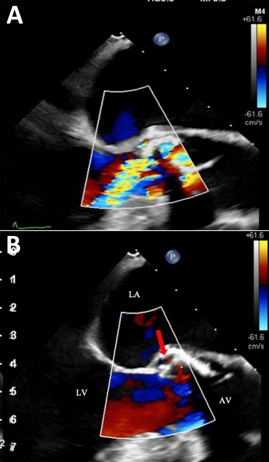
Color doppler echocardiography images from fatal case of subacute native aortic endocarditis, Geneva, Switzerland. A) Mid-esophageal long-axis view during diastole, showing moderate to severe aortic regurgitation. B) Mid-esophageal long-axis view with left ventricular chamber, aortic valve, and aortic root during systole. Red arrow indicates systolic flow with the pseudo-aneurysm. AV, open aortic valve; LA, left atrial; LV, left ventricle.

Figure 1. Color doppler echocardiography images from fatal case of subacute native aortic endocarditis, Geneva, Switzerland. A) Mid-esophageal long-axis view during diastole, showing moderate to severe aortic regurgitation. B) Mid-esophageal long-axis view with left ventricular chamber, aortic valve, and aortic root during systole. Red arrow indicates systolic flow with the pseudo-aneurysm. AV, open aortic valve; LA, left atrial; LV, left ventricle.

Main Article

1These authors are co–first authors.

Page created: May 31, 2025
Page updated: June 20, 2025
Page reviewed: June 20, 2025
The conclusions, findings, and opinions expressed by authors contributing to this journal do not necessarily reflect the official position of the U.S. Department of Health and Human Services, the Public Health Service, the Centers for Disease Control and Prevention, or the authors' affiliated institutions. Use of trade names is for identification only and does not imply endorsement by any of the groups named above.
file_external