Volume 31, Number 8—August 2025
Research Letter
Severe Fever with Thrombocytopenia Syndrome Acquired through Dog Bite, South Korea
Cite This Article
Citation for Media
Abstract
A veterinary technician in South Korea contracted severe fever with thrombocytopenia syndrome virus from a dog bite. Molecular evidence, including PCR sequencing, supports dog-to-human transmission. The case underscores the zoonotic risks posed by companion animals and highlights the importance of preventive measures.
Severe fever with thrombocytopenia syndrome (SFTS) is a zoonotic infectious disease caused by SFTS virus (Dabie bandavirus), primarily transmitted through tick bites (1). SFTS continues to spread across East Asia and poses a substantial public health threat; fatality rate in humans is ≈20% (1). Interspecies acquisition involving companion animals remains poorly understood; some reports suggest virus transmission from infected cats or dogs, but most lack definitive evidence such as documented bites (2–4). In South Korea, several canine SFTS cases have been reported (5). We describe a case of probable dog-to-human transmission of SFTSV through a bite, supported by molecular evidence.
A 23-year-old veterinary technician was transferred to Chonnam National University Hospital, a tertiary hospital in Gwangju, South Korea, after 6 days of fever. Laboratory findings showed leukopenia, thrombocytopenia, low C-reactive protein, and elevated liver enzymes and ferritin. The patient disclosed that a sick dog had bitten her right thumb 10 days before hospital admission; she had fed the dog wearing a mask but not gloves. The 7-mm wound had begun to heal after initial bleeding. The wound was rinsed under running water for <5 minutes before she arrived; hospital staff later applied antiseptic and administered a tetanus vaccine. Although the patient had no history of outdoor activity or known tick exposure, SFTS was suspected and subsequently confirmed by blood PCR. Her condition worsened on hospital day 2; we performed plasmapheresis on days 3 and 6 in the intensive care unit, leading to improvement.
The suspected source was a 4-year-old neutered male Pomeranian experiencing high fever, leukopenia, and thrombocytopenia, admitted to an animal hospital 10 days before the patient’s hospital admission. According to its owner, the dog had experienced 4 days of fever and anorexia; lethargy was first noted ≈22 days before hospital admission. After 2 weeks of supportive care, the dog fully recovered.
Figure
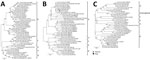
Figure. Phylogenetic analysis of SFTSV small (321 bp) (A), medium (477 bp) (B), and large (696 bp) (C) segments from human patient and dog, South Korea. Clustal X version 2.1 (...
To investigate potential dog-to-human transmission of SFTSV, we tested samples from the patient and the dog by reverse transcription PCR and immunofluorescent assay as previously described (5,6). To confirm the quantitative PCR results targeting the small segment by using the careGENE SFTS Virus RT-PCR kit (Wells Bio, https://www.wellsbio.net), we performed nested PCR. The dog’s saliva (collected on patient’s hospital day 4) showed a low level of SFTSV RNA (cycle threshold value 36.44), although no band was visible on nested PCR (Table). Sequencing of the nested PCR amplicon from blood samples revealed 99.6% identity in the medium segment and 100% in the large segment (Figure). Virus culture and sequencing of the small segment were unsuccessful.
Although the dog lived in an urban area, it was walked daily in nearby parks with dense vegetation. In the epidemiologic investigation, no ticks were found directly on the dog. We collected a total of 11 ticks from a suspected tick exposure site, a 94 m–high trail (34°58′15′′N, 127°33′51′′E) the dog and its owner frequently visited. The collected ticks included 6 adult female, 3 adult male, and 1 nymph Haemaphysalis longicornis ticks and 1 Ixodes granulatus nymph. All collected ticks tested negative for SFTSV.
A total of 43 persons, 3 household members with direct exposure to its saliva or bodily fluids and 40 veterinary staff, had contact with the dog. The Korea Disease Control and Prevention Agency recommended 2 weeks of symptom monitoring and PCR testing for those with direct contact. No PCR-positive or symptomatic cases were identified. None of the patient’s 31 hospital contacts experienced symptoms.
This case provides strong molecular evidence of dog-to-human SFTSV transmission from a bite. Unlike previous reports that relied solely on serologic findings (2,7), this case was supported by sequence identity and a documented bite. Although we could not isolate viable virus and the viral load in saliva was low, our findings suggest that canine saliva, particularly through dog bites, represents a potential transmission route, consistent with previous studies that detected SFTSV RNA in dog oral swab specimens (8) and isolated live virus at ≈106 RNA copies/mL concentration from cat saliva (9). A limitation of this study is the inability to culture the virus, likely caused by delays in specimen collection. In addition, the absence of early saliva samples limits definitive confirmation of bite-mediated transmission, although the epidemiologic and molecular findings strongly support this route. Serologic testing was not performed for contacts, and asymptomatic cases may have gone undetected.
This case underscores the importance of personal protective equipment and infection control in both veterinary and human healthcare settings to prevent zoonotic transmission. Prompt wound care after animal bite or scratches, including washing with soap and running water for >20 minutes, can reduce the risk for infections such as B virus or rabies (10). Although the effectiveness of this approach against SFTS is unproven, the same principle may reduce the risk for other viral infections transmitted through animal bites or saliva exposure.
In conclusion, this case emphasizes the risk for SFTSV transmission not only via tick bites but also directly through bites from infected dogs. Enhanced awareness and preventive strategies in both veterinary and human healthcare settings are critical to mitigating the risks of SFTS.
Dr. Kim is an infectious disease specialist and an associate professor at Chonnam National University Hospital in Gwangju, South Korea. Her primary interests are zoonoses and hospital infection control.
Acknowledgments
We thank the patient and the animal hospital staff for granting permission to report their clinical symptoms and disease courses.
The patient provided written informed consent for publication. The Institutional Review Board of Chonnam National University granted ethics approval for this study (IRB no. CNUH-2022-032).
The Chonnam National University Hospital Biomedical Research Institute supported this study (grant no. BCR124055).
References
- Cui H, Shen S, Chen L, Fan Z, Wen Q, Xing Y, et al. Global epidemiology of severe fever with thrombocytopenia syndrome virus in human and animals: a systematic review and meta-analysis. Lancet Reg Health West Pac. 2024;48:
101133 . DOIPubMedGoogle Scholar - Oshima H, Okumura H, Maeda K, Ishijima K, Yoshikawa T, Kurosu T, et al. A patient with severe fever with thrombocytopenia syndrome (SFTS) infected from a sick dog with SFTS virus infection. Jpn J Infect Dis. 2022;75:423–6. DOIPubMedGoogle Scholar
- Yamanaka A, Kirino Y, Fujimoto S, Ueda N, Himeji D, Miura M, et al. Direct transmission of severe fever with thrombocytopenia syndrome virus from domestic cat to veterinary personnel. Emerg Infect Dis. 2020;26:2994–8. DOIPubMedGoogle Scholar
- Kobayashi Y, Kato H, Yamagishi T, Shimada T, Matsui T, Yoshikawa T, et al.; SFTS Epidemiological Research Group Japan. Severe fever with thrombocytopenia syndrome, Japan, 2013–2017. Emerg Infect Dis. 2020;26:692–9. DOIPubMedGoogle Scholar
- Han SW, Kang JG, Byeon AR, Cho YK, Choi KS, Chae JS. Severe fever with thrombocytopenia syndrome in canines from the Republic of Korea. Ticks Tick Borne Dis. 2020;11:
101454 . DOIPubMedGoogle Scholar - Kim CM, Han MA, Yun NR, Bang MS, Lee YM, Lee B, et al. Seroprevalence of severe fever with thrombocytopenia syndrome using specimens from the Korea National Health & Nutrition Examination Survey. PLoS Negl Trop Dis. 2023;17:
e0011097 . DOIPubMedGoogle Scholar - Tsuru M, Suzuki T, Murakami T, Matsui K, Maeda Y, Yoshikawa T, et al. Pathological characteristics of a patient with severe fever with thrombocytopenia syndrome (SFTS) infected with SFTS virus through a sick cat’s bite. Viruses. 2021;13:204. DOIPubMedGoogle Scholar
- Ishijima K, Tatemoto K, Park E, Kimura M, Fujita O, Taira M, et al. Lethal disease in dogs naturally infected with severe fever with thrombocytopenia syndrome virus. Viruses. 2022;14:1963. DOIPubMedGoogle Scholar
- Park ES, Shimojima M, Nagata N, Ami Y, Yoshikawa T, Iwata-Yoshikawa N, et al. Severe Fever with Thrombocytopenia Syndrome Phlebovirus causes lethal viral hemorrhagic fever in cats. Sci Rep. 2019;9:11990. DOIPubMedGoogle Scholar
- Centers for Disease Control and Prevention. CDC yellow book 2026: health information for international travel. Oxford: Oxford University Press; 2025.
Figure
Table
Cite This ArticleOriginal Publication Date: July 18, 2025
1These first authors contributed equally to this article.
2These senior authors contributed equally to this article.
Table of Contents – Volume 31, Number 8—August 2025
| EID Search Options |
|---|
|
|
|
|
|
|
Please use the form below to submit correspondence to the authors or contact them at the following address:
Kyung-Hwa Park, Department of Infectious Diseases, Chonnam National University Medical School, 42 Jaebongro, Dong-gu, Gwangju 61469, South Korea
Top