Volume 31, Number 9—September 2025
Dispatch
Gastric Submucosal Tumor in Patient Infected with Dioctophyme renale Roundworm, South Korea, 2024
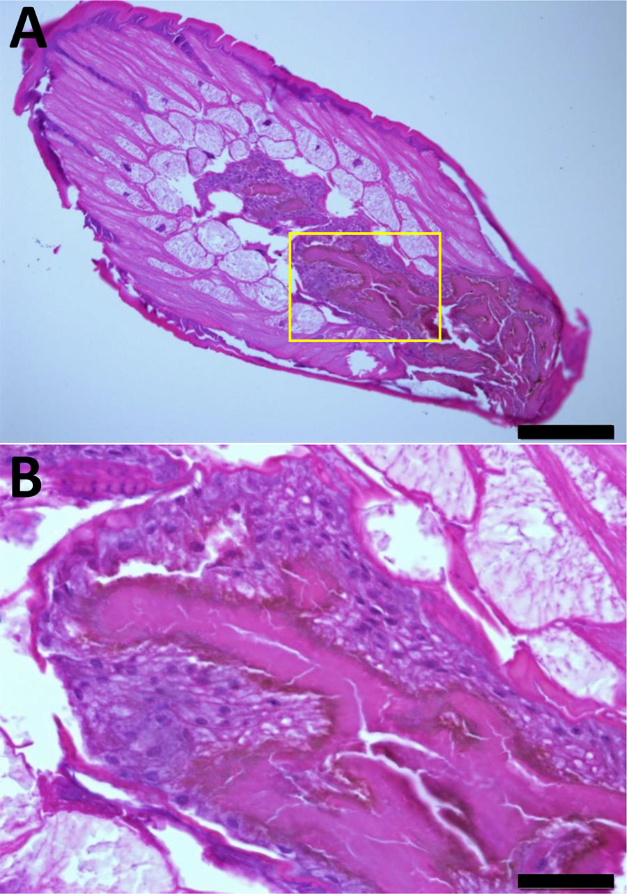
Figure 2

Figure 2. Dioctophym renale larva stained with hematoxylin and eosin in a study of gastric submucosal tumor in patient infected with D. renale roundworm, South Korea, 2024. A) Cross-section displays long subcuticular polymyarian musculature and characteristic intestines. Spines were not observed on the surface of the body. The intestinal epithelium consists of multilayered cuboidal cells with relatively large nuclei. Numerous dark brown granules (hematoxylin and eosin stain) are visible along the luminal border and are covered with microvilli. Scale bar indicates 200 μm. B) Higher magnification of the specimen shown in panel A (yellow box), providing a closer view of the characteristic intestine. These images suggested the presence of D. renale L3 larva. Scale bar indicates 2050 μm.
1These authors contributed equally to this study.