Volume 31, Number 9—September 2025
Dispatch
Emergence of Autochthonous Leishmania (Mundinia) martiniquensis Infections in Horses, Czech Republic and Austria, 2019–2023
David Modrý , Edmund K. Hainisch, Hans-Peter Fuehrer, Edwin Kniha, Maria Sophia Unterköfler, Jovana Sádlová, Petr Jahn, Kristína Řeháková, Kamil Sedlák, and Jan Votýpka
Figure 1
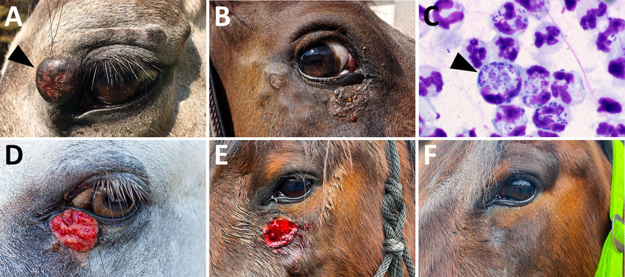
Figure 1. Cutaneous lesions during initial clinical examination and detection of Leishmania amastigotes from study of autochthonous Leishmania (Mundinia) martiniquensis infections in horses, Czech Republic and Austria, 2019–2023. A) Periorbital nodular lesions from case 1 (L. martiniquensis was cultured from a fine needle aspiration biopsy of the largest lesion, indicated by arrowhead); B) periorbital lesions in case 2; C) Leishmania amastigotes (indicated by arrowhead) in a stained smear from sample from case 1; D) lower eyelid lesion in case 3 (image by Christian Bernkopf); E) facial lesions in case 4 at the time of Leishmania detection; F) photograph of case 4 horse showing no recurrence 27 months later.
Page created: July 11, 2025
Page updated: August 26, 2025
Page reviewed: August 26, 2025
The conclusions, findings, and opinions expressed by authors contributing to this journal do not necessarily reflect the official position of the U.S. Department of Health and Human Services, the Public Health Service, the Centers for Disease Control and Prevention, or the authors' affiliated institutions. Use of trade names is for identification only and does not imply endorsement by any of the groups named above.