Skip directly to site content Skip directly to page options Skip directly to A-Z link Skip directly to A-Z link Skip directly to A-Z link

Volume 31, Number 9—September 2025

Research

Attachment Patterns of Avian Influenza H5 Clade 2.3.4.4b Virus in Respiratory Tracts of Marine Mammals, North Atlantic Ocean

Syriam Sooksawasdi Na Ayudhya, Lonneke Leijten, Willemijn F. Rijnink, Monique I. Spronken, Thijs Kuiken, Lisa Bauer1, and Debby van Riel1Comments to Author 
Author affiliation: Prince of Songkla University, Faculty of Veterinary Science, Songkhla, Thailand (S. Sooksawasdi Na Ayudhya); Erasmus University Rotterdam, Erasmus Medical Center, Rotterdam, the Netherlands (S. Sooksawasdi Na Ayudhya, L. Leijten, W.F. Rijnink, M.I. Spronken, T. Kuiken, L. Bauer, D. van Riel)

Main Article

Figure 1

Attachment of influenza A viruses to the olfactory mucosa and respiratory mucosa of seals in study of attachment patterns of avian influenza H5 clade 2.3.4.4b virus in respiratory tracts of marine mammals, North Atlantic Ocean. Hematoxylin and eosin stain (red) shows attachment of human seasonal influenza A virus H32003 and avian influenza A viruses H5ΔMBCS2005 and H5ΔMBCS2022 to the olfactory (A) and respiratory (B) mucosa in the nasal turbinate of harbor seals (Phoca vitulina) and gray seals (Halichoerus grypus). Photos were taken at high magnification (×1,000) of the apical side of the mucosa; for this reason, Bowman’s glands in the submucosa are not represented. H32003, A/Netherlands/213/2003 (H3N2); H5ΔMBCS2005, A/Indonesia/05/2005 (H5N1); H5ΔMBCS2022, A/Caspian gull/Netherlands/1/2022 (H5N1).

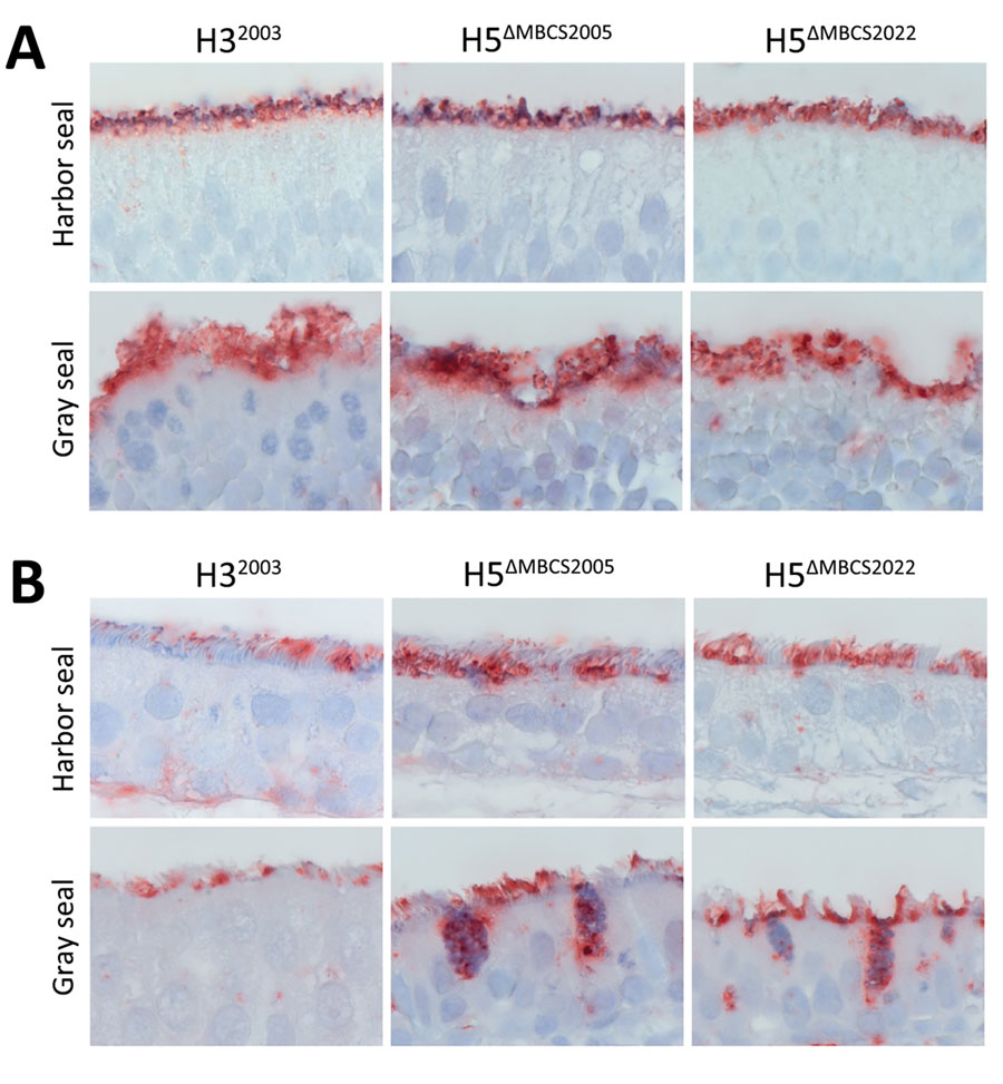
Figure 1. Attachment of influenza A viruses to the olfactory mucosa and respiratory mucosa of seals in study of attachment patterns of avian influenza H5 clade 2.3.4.4b virus in respiratory tracts of marine mammals, North Atlantic Ocean. Hematoxylin and eosin stain (red) shows attachment of human seasonal influenza A virus H32003 and avian influenza A viruses H5ΔMBCS2005 and H5ΔMBCS2022 to the olfactory (A) and respiratory (B) mucosa in the nasal turbinate of harbor seals (Phoca vitulina) and gray seals (Halichoerus grypus). Photos were taken at high magnification (×1,000) of the apical side of the mucosa; for this reason, Bowman’s glands in the submucosa are not represented. H32003, A/Netherlands/213/2003 (H3N2); H5ΔMBCS2005, A/Indonesia/05/2005 (H5N1); H5ΔMBCS2022, A/Caspian gull/Netherlands/1/2022 (H5N1).

Main Article

1Current affiliation: Prince of Songkla University, Songkhla, Thailand.

Page created: July 23, 2025
Page updated: September 17, 2025
Page reviewed: September 17, 2025
The conclusions, findings, and opinions expressed by authors contributing to this journal do not necessarily reflect the official position of the U.S. Department of Health and Human Services, the Public Health Service, the Centers for Disease Control and Prevention, or the authors' affiliated institutions. Use of trade names is for identification only and does not imply endorsement by any of the groups named above.
file_external