Volume 32, Number 1—January 2026
Synopsis
Pulmonary Histoplasmosis, Taiwan, 1997–2024
Ting-Wei Kao, Shang-Chen Yang, Hsiang-Wei Hu, Yu-Tsung Huang, Chin-Chung Shu , and Wang-Huei Sheng
Figure 1
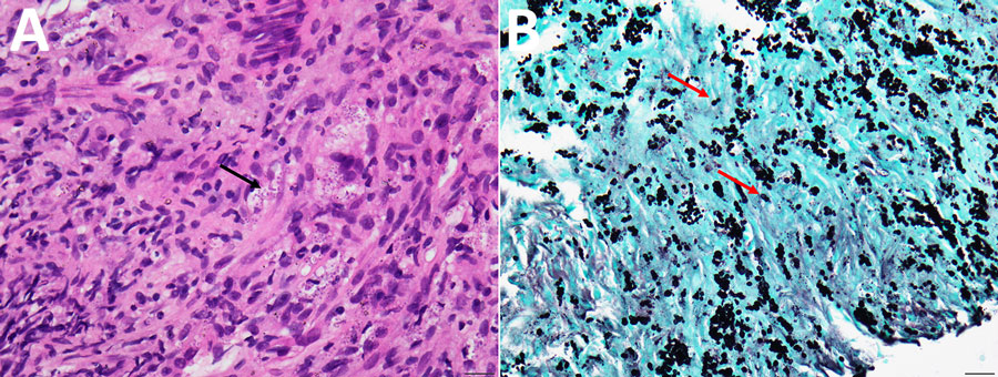
Figure 1. Representative histopathologic findings of pulmonary histoplasmosis in Taiwan, 1997–2024. A) Hematoxylin and eosin stain delineates numerous tiny ovoid yeasts within macrophages; occasional narrow-based budding indicated (black arrow). B) Grocott's methenamine silver stain shows yeasts with narrow-based budding (red arrows). Scale bars represent 20 µm.
Page created: December 06, 2025
Page updated: January 27, 2026
Page reviewed: January 27, 2026
The conclusions, findings, and opinions expressed by authors contributing to this journal do not necessarily reflect the official position of the U.S. Department of Health and Human Services, the Public Health Service, the Centers for Disease Control and Prevention, or the authors' affiliated institutions. Use of trade names is for identification only and does not imply endorsement by any of the groups named above.