Skip directly to site content Skip directly to page options Skip directly to A-Z link Skip directly to A-Z link Skip directly to A-Z link

Disclaimer: Early release articles are not considered as final versions. Any changes will be reflected in the online version in the month the article is officially released.

Volume 32, Number 10—October 2026

Research

Experimental Highly Pathogenic Avian Influenza A(H5N1) Clade 2.3.4.4b Virus Infection in Alpacas, 2026

Jacob SchönComments to Author , Angele Breithaupt, Nico Joel Halwe, Maxi Hertel, Ann Kathrin Ahrens, Andrea Aebischer, Donata Hoffmann, and Martin Beer
Author affiliation: Institute of Diagnostic Virology, Friedrich-Loeffler-Institut, Greifswald, Germany (J. Schön, N.J. Halwe, A.K. Ahrens, A. Aebischer, D. Hoffman, M. Beer); Department of Experimental Animal Facilities and Biorisk Management, Friedrich-Loeffler-Institut, Greifswald (A. Breithaupt, M. Hertel)

Main Article

Figure 1

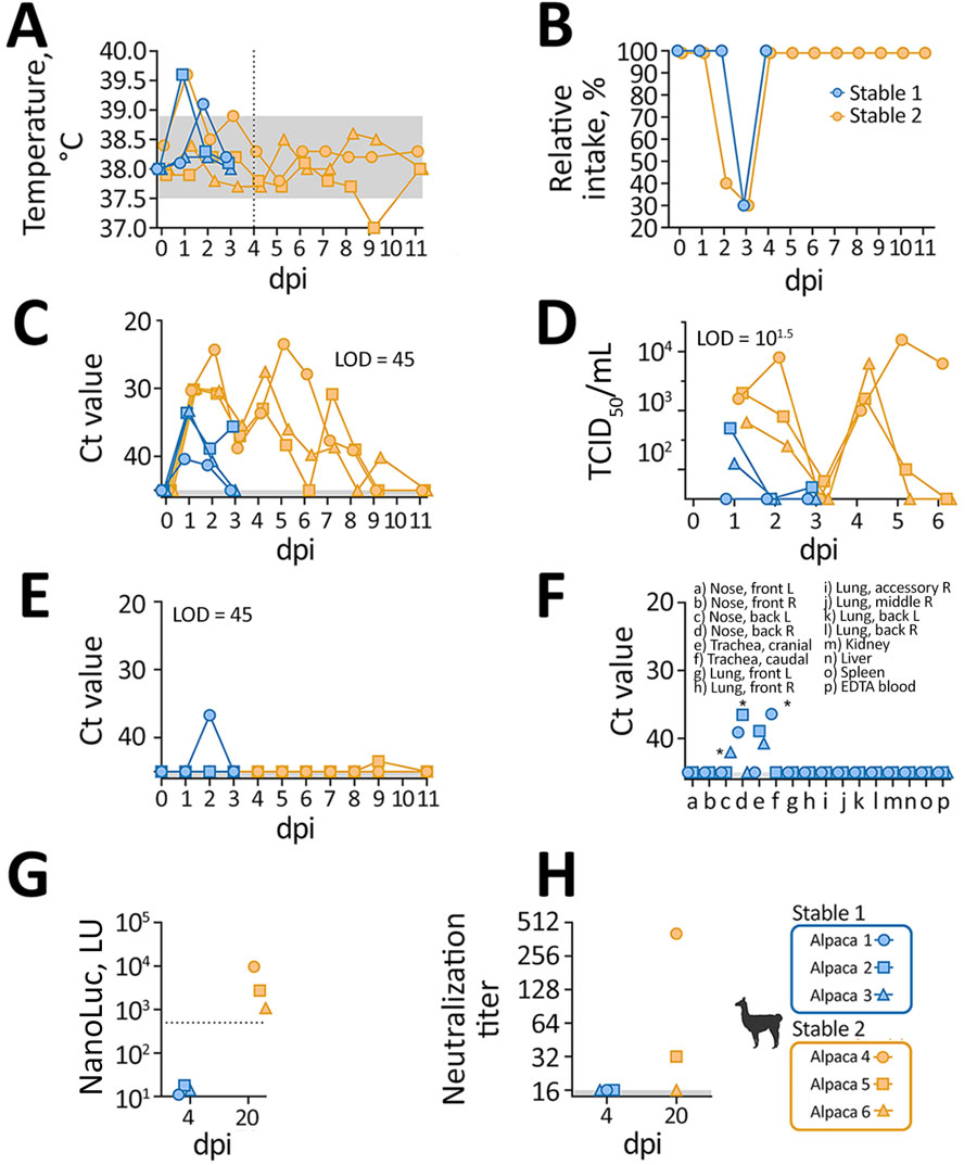
Clinical and laboratory findings from experimental highly pathogenic avian influenza A(H5N1) clade 2.3.4.4b virus infection in alpacas, 2026. Stable 1 alpacas were sampled before euthanasia at 4 dpi; stable 2 alpacas were sampled for 20 dpi before euthanasia. A, B) Clinical signs; C–H) laboratory findings. A) Body temperature; shading indicates physiologic range; dotted vertical line indicates dpi that stable 1 alpacas were euthanized. B) Percentage of typical feed intake. C) Quantitative reverse transcription PCR (qRT-PCR) results on nasal swab samples showing H5N1 viral genome was detected until 7 dpi. Shading indicates LOD.  D) Nasal swab sample infectious titers. E) qRT-PCR results on rectal swab samples. Shading indicates LOD. F) qRT-PCR results on tissue samples. Of note, only nose and trachea samples were qRT-PCR–positive as evaluated by endpoint dilution assays for quantification of infectious virus. Asterisks indicate samples that tested positive for infectivity by endpoint dilution at 101.88, 101.82, and 101.62 TCID50/mL. G) ELISA specific for H5 antibodies, confirming positivity of samples collected at 20 dpi. H) Neutralization titers determined by using a live virus neutralization test against 100 TCID50, with the endpoint defined as complete protection from cytopathic effect. Titers are expressed as reciprocal serum dilution, confirming measurable neutralizing capacity for 2 of the samples that tested positive by ELISA. dpi, days postinfection; L, left; LOD, limit of detection; LU, light unit; NanoLuc, nanoluciferase; R, right; TCID50, 50% tissue infective dose.

Figure 1. Clinical and laboratory findings from experimental highly pathogenic avian influenza A(H5N1) clade 2.3.4.4b virus infection in alpacas, 2026. Stable 1 alpacas were sampled before euthanasia at 4 dpi; stable 2 alpacas were sampled for 20 dpi before euthanasia. A, B) Clinical signs; C–H) laboratory findings. A) Body temperature; shading indicates physiologic range; dotted vertical line indicates dpi that stable 1 alpacas were euthanized. B) Percentage of typical feed intake. C) Quantitative reverse transcription PCR (qRT-PCR) results on nasal swab samples showing H5N1 viral genome was detected until 7 dpi. Shading indicates LOD. D) Nasal swab sample infectious titers. E) qRT-PCR results on rectal swab samples. Shading indicates LOD. F) qRT-PCR results on tissue samples. Of note, only nose and trachea samples were qRT-PCR–positive as evaluated by endpoint dilution assays for quantification of infectious virus. Asterisks indicate samples that tested positive for infectivity by endpoint dilution at 101.88, 101.82, and 101.62 TCID50/mL. G) ELISA specific for H5 antibodies, confirming positivity of samples collected at 20 dpi. H) Neutralization titers determined by using a live virus neutralization test against 100 TCID50, with the endpoint defined as complete protection from cytopathic effect. Titers are expressed as reciprocal serum dilution, confirming measurable neutralizing capacity for 2 of the samples that tested positive by ELISA. dpi, days postinfection; L, left; LOD, limit of detection; LU, light unit; NanoLuc, nanoluciferase; R, right; TCID50, 50% tissue infective dose.

Main Article

Page created: August 05, 2026
Page updated: September 16, 2026
Page reviewed: September 16, 2026
The conclusions, findings, and opinions expressed by authors contributing to this journal do not necessarily reflect the official position of the U.S. Department of Health and Human Services, the Public Health Service, the Centers for Disease Control and Prevention, or the authors' affiliated institutions. Use of trade names is for identification only and does not imply endorsement by any of the groups named above.
file_external