Skip directly to site content Skip directly to page options Skip directly to A-Z link Skip directly to A-Z link Skip directly to A-Z link

Disclaimer: Early release articles are not considered as final versions. Any changes will be reflected in the online version in the month the article is officially released.

Volume 32, Number 11—November 2026

Research Letter

Disseminated Trimethoprim/Sulfamethoxazole–Resistant Nocardiosis in Renal Transplant Recipient, India

Sandeep Rao Kordcal, Md Tariq Maula, Vivek Malleshappa Suguna, Adil Rashid Khan, Rahul Ranjan, Stanzin Spalkit, Aruna Nambirajan, Gagandeep Singh, Neeraj Nischal, Immaculata Xess, and Manish SonejaComments to Author 
Author affiliation: All India Institute of Medical Sciences, New Delhi, India

Main Article

Figure 1

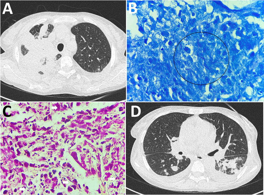
Histopathologic and radiologic findings in renal transplant recipient with disseminated trimethoprim/sulfamethoxazole–resistant nocardiosis, India. A) High-resolution computed tomography of the chest before treatment, showing right-upper-lobe consolidation with adjacent passive atelectasis and a left pleural effusion. B) Lung biopsy, Fite stained, showing weakly acid-fast, branching, beaded filamentous bacteria (circle). Original magnification ×1,000. C) Lung biopsy, hematoxylin and eosin stained, showing suppurative inflammation (arrows). Original magnification ×400. D) High-resolution computed tomography of the chest 6 months after treatment, showing resolution of the consolidation with residual pulmonary fibrosis.

Figure 1. Histopathologic and radiologic findings in renal transplant recipient with disseminated trimethoprim/sulfamethoxazole–resistant nocardiosis, India. A) High-resolution computed tomography of the chest before treatment, showing right-upper-lobe consolidation with adjacent passive atelectasis and a left pleural effusion. B) Lung biopsy, Fite stained, showing weakly acid-fast, branching, beaded filamentous bacteria (circle). Original magnification ×1,000. C) Lung biopsy, hematoxylin and eosin stained, showing suppurative inflammation (arrows). Original magnification ×400. D) High-resolution computed tomography of the chest 6 months after treatment, showing resolution of the consolidation with residual pulmonary fibrosis.

Main Article

Page created: September 11, 2026
Page updated: October 15, 2026
Page reviewed: October 15, 2026
The conclusions, findings, and opinions expressed by authors contributing to this journal do not necessarily reflect the official position of the U.S. Department of Health and Human Services, the Public Health Service, the Centers for Disease Control and Prevention, or the authors' affiliated institutions. Use of trade names is for identification only and does not imply endorsement by any of the groups named above.
file_external