Cutaneous Paraconiothyrium cyclothyrioides Infection in Lung Transplant Recipient, Georgia, USA
Carolyn Mackey

, Stephanie Thomas, Lucy S. Witt, Elizabeth Sajewski, Shawn R. Lockhart, Stephanie M. Pouch, Justin Cheeley, Jamie B. MacKelfresh, and Jeremy A.W. Gold
Author affiliation: Georgia Emerging Infections Program, Atlanta, Georgia, USA (C. Mackey, S. Thomas, L.S. Witt, S.M. Pouch); Atlanta Veterans Affairs Health System, Decatur, Georgia, USA (C. Mackey, S. Thomas, L.S. Witt, S.M. Pouch); Emory University School of Medicine, Atlanta (C. Mackey, S. Thomas, L.S. Witt, S.M. Pouch, J. Cheeley, J.B. MacKelfresh); Centers for Diseases Control and Prevention, Atlanta (E. Sajewski, S.R. Lockhart, J.A.W. Gold)
Main Article
Figure 2
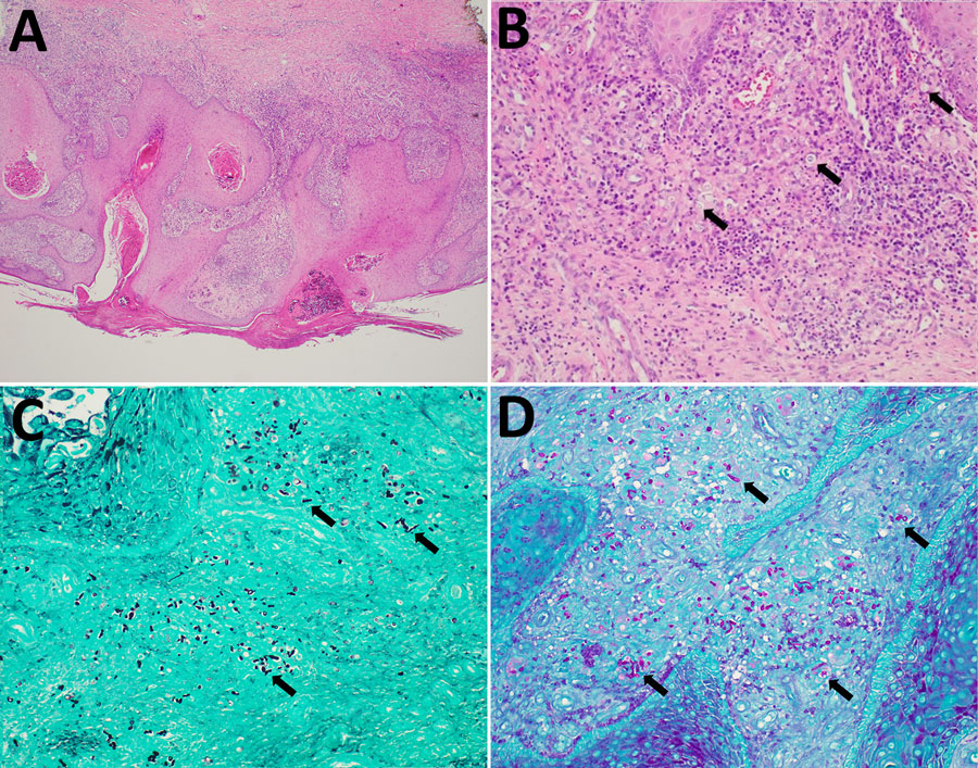
Figure 2. Histologic (A, B) and histochemical (C, D) findings from case of cutaneous Paraconiothyrium cyclothyrioides in lung transplant recipient, Georgia, USA. A) Skin biopsy from right shin revealed psuedoepitheliomatous hyperplasia of the epidermis overlying a mixed dermal inflammatory infiltrate. Hematoxylin and eosin stain; original magnification ×40. B) Polymorphic fungal elements (arrows) seen within the superficial dermis. Hematoxylin and eosin stain; original magnification ×200. C) Numerous fungal hyphal elements and yeast-like forms (arrows) highlighted within the dermis. Grocott methenamine silver stain; original magnification ×200. D) Fungal hyphal elements and yeast-like forms (arrows) noted by Periodic acid–Schiff stain; original magnification ×200.
Main Article
Page created: February 27, 2026
Page updated: March 20, 2026
Page reviewed: March 20, 2026
The conclusions, findings, and opinions expressed by authors contributing to this journal do not necessarily reflect the official position of the U.S. Department of Health and Human Services, the Public Health Service, the Centers for Disease Control and Prevention, or the authors' affiliated institutions. Use of trade names is for identification only and does not imply endorsement by any of the groups named above.