Volume 32, Number 4—April 2026
Dispatch
Disseminated Acanthamoeba Infection with Necrotic Skin Lesions and Granulomatous Vasculitis, United States
Maria Koshy , Carrie Flynn, Marat Kribis, Jennifer McNiff, Matthew Grant, and Shana Elizabeth Gleeson
Figure 4
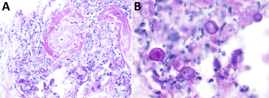
Figure 4. Periodic acid-Schiff positive large histiocytoid cells near the vessel, from a patient with disseminated Acanthamoeba infection with necrotic skin lesions and granulomatous vasculitis. Cells are potential amoebic trophozoites. A) Original magnification ×400; B) Original magnification ×600.
Page created: March 12, 2026
Page updated: April 15, 2026
Page reviewed: April 15, 2026
The conclusions, findings, and opinions expressed by authors contributing to this journal do not necessarily reflect the official position of the U.S. Department of Health and Human Services, the Public Health Service, the Centers for Disease Control and Prevention, or the authors' affiliated institutions. Use of trade names is for identification only and does not imply endorsement by any of the groups named above.