Skip directly to site content Skip directly to page options Skip directly to A-Z link Skip directly to A-Z link Skip directly to A-Z link

Disclaimer: Early release articles are not considered as final versions. Any changes will be reflected in the online version in the month the article is officially released.

Volume 32, Number 5—May 2026

Dispatch

Replication Efficiency of Contemporary Highly Pathogenic Avian Influenza A(H5N1) Virus Isolates in Human Nasal Epithelium Model

Meaghan Flagg1, Christopher J. Winski1, Bridget G. Brackney, Tessa R. Lutterman, Johan A. Ortiz-Morales2, Brandi N. Williamson, and Emmie de WitComments to Author 
Author affiliation: Author affiliation: National Institute of Allergy and Infectious Diseases, National Institutes of Health, Hamilton, Montana, USA

Main Article

Figure 3

Induction of proinflammatory cytokines in human nasal epithelium infected with seasonal influenza A and highly pathogenic avian influenza (HPAI) A(H5N1) viruses in study of replication efficiency of contemporary HPAI H5N1 virus isolates in human nasal epithelium model. We inoculated Mattek EpiNasal cultures (https://www.mattek.com) with a multiplicity of infection of 0.1 50% tissue culture infectious dose per cell. RNA was extracted from cells at 0, 8, 24, 48, 72, and 96 hours postinoculation. We ran quantitative reverse transcription PCR using primers (Integrated DNA Technologies, https://www.idtdna.com) to detect interleukin 6 (A), tumor necrosis factor α (B), and interleukin 1β (C) for historical and clade 2.3.4.4b HPAI H5N1 genotype B3.13, B3.6, and D1.1 strains. Lines indicate medians; shading indicates 95% CIs. We normalized data to internal controls (ACTB and GAPDH) and calculated fold change relative to timepoint-matched mock-infected controls. Fold change is reported for 3 biologic replicates. We performed statistical analysis using 2-way analysis of variance with Dunnett posttest. p values are shown by asterisks in colors matching isolates: *p<0.05; **p<0.01; ***p<0.001; ****p<0.0001.

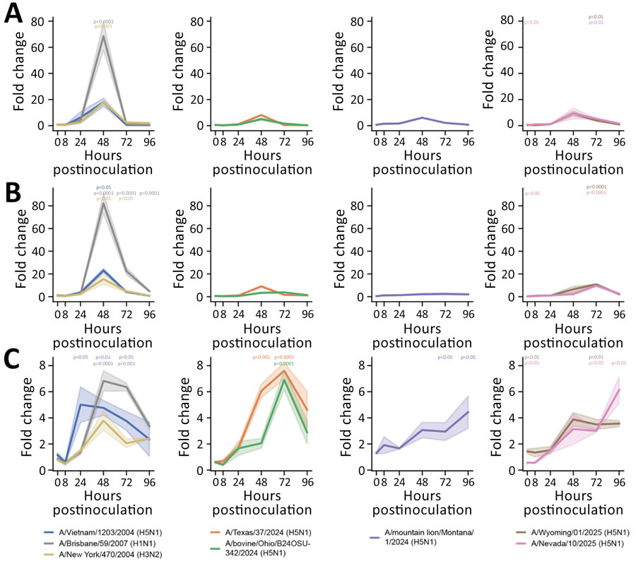
Figure 3. Induction of proinflammatory cytokines in human nasal epithelium infected with seasonal influenza A and highly pathogenic avian influenza (HPAI) A(H5N1) viruses in study of replication efficiency of contemporary HPAI H5N1 virus isolates in human nasal epithelium model. We inoculated Mattek EpiNasal cultures (https://www.mattek.com) with a multiplicity of infection of 0.1 50% tissue culture infectious dose per cell. RNA was extracted from cells at 0, 8, 24, 48, 72, and 96 hours postinoculation. We ran quantitative reverse transcription PCR using primers (Integrated DNA Technologies, https://www.idtdna.com) to detect interleukin 6 (A), tumor necrosis factor α (B), and interleukin 1β (C) for historical and clade 2.3.4.4b HPAI H5N1 genotype B3.13, B3.6, and D1.1 strains. Lines indicate medians; shading indicates 95% CIs. We normalized data to internal controls (ACTB and GAPDH) and calculated fold change relative to timepoint-matched mock-infected controls. Fold change is reported for 3 biologic replicates. We performed statistical analysis using 2-way analysis of variance with Dunnett posttest. p values are shown by asterisks in colors matching isolates: *p<0.05; **p<0.01; ***p<0.001; ****p<0.0001.

Main Article

1These authors contributed equally to this article.

2Current affiliation: LSU Health, Shreveport, Louisiana, USA.

Page created: April 14, 2026
Page updated: May 01, 2026
Page reviewed: May 01, 2026
The conclusions, findings, and opinions expressed by authors contributing to this journal do not necessarily reflect the official position of the U.S. Department of Health and Human Services, the Public Health Service, the Centers for Disease Control and Prevention, or the authors' affiliated institutions. Use of trade names is for identification only and does not imply endorsement by any of the groups named above.
file_external