Skip directly to site content Skip directly to page options Skip directly to A-Z link Skip directly to A-Z link Skip directly to A-Z link

Disclaimer: Early release articles are not considered as final versions. Any changes will be reflected in the online version in the month the article is officially released.

Volume 32, Number 5—May 2026

Dispatch

Replication Efficiency of Contemporary Highly Pathogenic Avian Influenza A(H5N1) Virus Isolates in Human Nasal Epithelium Model

Meaghan Flagg1, Christopher J. Winski1, Bridget G. Brackney, Tessa R. Lutterman, Johan A. Ortiz-Morales2, Brandi N. Williamson, and Emmie de WitComments to Author 
Author affiliation: Author affiliation: National Institute of Allergy and Infectious Diseases, National Institutes of Health, Hamilton, Montana, USA

Main Article

Figure 1

Replication of seasonal and highly pathogenic avian influenza (HPAI) A viruses in human nasal epithelium cultures in study of replication efficiency of contemporary HPAI H5N1 virus isolates in human nasal epithelium model. A–D) Virus replication in historical (A) and HPAI H5N1 genotype clade 2.3.4.4b B3.13 (B), B3.6 (C), and D1.1 (D) virus strains. We inoculated Mattek EpiNasal cultures (https://www.mattek.com) with a multiplicity of infection of 0.1 TCID50 per cell. We harvested apical supernatant at 0, 8, 24, 48, 72, and 96 hours postinoculation and titered on MDCK cells. Data points and error bars represent the geometric mean +SD of 3 biologic replicates from a single donor; dashed line indicates lower limit of detection. E) Area under the curve of data from 0–96 hours postinoculation as shown in panels A–D. Error bars indicate SD. F) Statistical analysis of data in panel E performed using 1-way analysis of variance with multiple comparisons (Tukey). NS, not significant; TCID50, 50% tissue culture infectious dose.

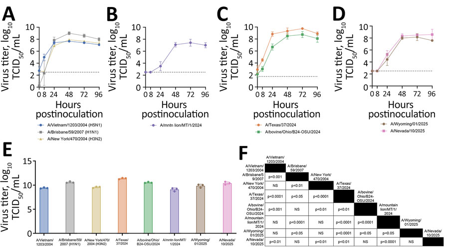
Figure 1. Replication of seasonal and highly pathogenic avian influenza (HPAI) A viruses in human nasal epithelium cultures in study of replication efficiency of contemporary HPAI H5N1 virus isolates in human nasal epithelium model. A–D) Virus replication in historical (A) and HPAI H5N1 genotype clade 2.3.4.4b B3.13 (B), B3.6 (C), and D1.1 (D) virus strains. We inoculated Mattek EpiNasal cultures (https://www.mattek.com) with a multiplicity of infection of 0.1 TCID50 per cell. We harvested apical supernatant at 0, 8, 24, 48, 72, and 96 hours postinoculation and titered on MDCK cells. Data points and error bars represent the geometric mean +SD of 3 biologic replicates from a single donor; dashed line indicates lower limit of detection. E) Area under the curve of data from 0–96 hours postinoculation as shown in panels A–D. Error bars indicate SD. F) Statistical analysis of data in panel E performed using 1-way analysis of variance with multiple comparisons (Tukey). NS, not significant; TCID50, 50% tissue culture infectious dose.

Main Article

1These authors contributed equally to this article.

2Current affiliation: LSU Health, Shreveport, Louisiana, USA.

Page created: April 14, 2026
Page updated: May 01, 2026
Page reviewed: May 01, 2026
The conclusions, findings, and opinions expressed by authors contributing to this journal do not necessarily reflect the official position of the U.S. Department of Health and Human Services, the Public Health Service, the Centers for Disease Control and Prevention, or the authors' affiliated institutions. Use of trade names is for identification only and does not imply endorsement by any of the groups named above.
file_external