Volume 32, Number 5—May 2026
Dispatch
Highly Pathogenic Avian Influenza A(H5N1) Clade 2.3.4.4b Virus and Mass Mortality in Eurasian Cranes, Germany, 2025
Anne Günther, Christof Herrmann, Julia Sehl-Ewert, Simon Piro, Ann Kathrin Ahrens, Sten Calvelage, Anne Pohlmann, Martin Beer, and Timm Harder
Figure 2
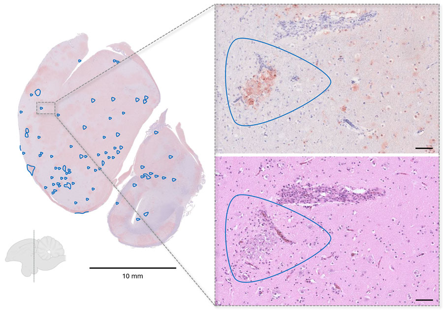
Figure 2. Representative histopathologic findings in influenza A(H5N1)–infected cranes from study of highly pathogenic avian influenza A(H5N1) clade 2.3.4.4b virus causing mass mortality in cranes, Germany, 2025. Immunohistochemistry of a coronal brain section at the level of the nidopallium demonstrating widespread viral antigen detection (brown staining) with only limited inflammatory and necrotic changes (blue outlines). Callout images at right show the corresponding region in consecutive sections, with abundant antigen-positive cells in immunohistochemistry (top) but only a small necrotic focus in the matching hematoxylin and eosin–stained section (bottom). Scale bars in callout images represent 50 μm.
Page created: April 14, 2026
Page updated: May 15, 2026
Page reviewed: May 15, 2026
The conclusions, findings, and opinions expressed by authors contributing to this journal do not necessarily reflect the official position of the U.S. Department of Health and Human Services, the Public Health Service, the Centers for Disease Control and Prevention, or the authors' affiliated institutions. Use of trade names is for identification only and does not imply endorsement by any of the groups named above.