Disclaimer: Early release articles are not considered as final versions. Any changes will be reflected in the online version in the month the article is officially released.
Volume 32, Number 7—July 2026
Research Letter
New World Ocular Dirofilariasis Caused by Dirofilaria repens Infection, United States
Suggested citation for this article
Abstract
We describe an infection caused by Dirofilaria repens nematodes in California, USA. A firm nodule developed after an insect bite on a patient’s eyelid. Excision with morphologic and molecular analysis confirmed D. repens infection. Our findings confirm the necessity of both molecular and histological studies to identify nematode infections.
Dirofilariasis is caused by Dirofilaria (family Onchocercidae) nematodes. Old World infections are commonly caused by Dirofilaria repens nematodes. New World infections are generally caused by species other than D. repens, such as D. immitis, D. tenuis, D. subdermata, D. striata, and D. ursi (1). Canids, felids, and raccoons are the definitive hosts for most zoonotic infections, and mosquitoes serve as intermediate vectors. In humans, ocular dirofilariasis, which includes eyelid, subconjunctival, orbital, and intraocular infections, accounts for <35% of all cases (2). The eyelid and orbit are the sites of ≈42% of ocular dirofilariasis cases (3). The species causing ocular dirofilariasis have distinct geographic associations (3). We describe ocular dirofilariasis caused by D. repens nematodes in California, USA.
Figure 1
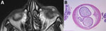
Figure 1. Images from patient with ocular dirofilariasis caused by Dirofilaria repensin California, USA. A) Axial plane of T-2–weighted orbital magnetic resonance imaging revealing a well circumscribed cystic lesion in...
A 74-year-old man from California was bitten by an insect on his left lower eyelid. Initially, he experienced transient pain, swelling, and weeping at the wound. Six weeks later, his dermatologist noted an 8-mm diameter, firm, nontender subcutaneous nodule at this site. The patient had no medical history or recent travel of note. After referral to an ophthalmologist, magnetic resonance imaging of the orbits confirmed a well-circumscribed cystic lesion on the eyelid (Figure 1, panel A). The mass persisted for 5 months, and an excisional biopsy was performed. The mass was dissected from closely adherent surrounding tissue and submitted to pathology in formalin. The patient was asymptomatic 6 months after the surgery.
The tissue was processed routinely and sectioned after paraffin embedding. Microscopy revealed a parasite surrounded by fibrosis with marked chronic inflammation. The cross section of the parasite revealed features of a nematode consistent with Dirofilaria sp. (Figure 1, panel B).
Figure 2
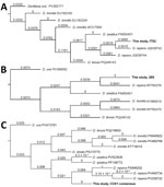
Figure 2. Dirofilaria repensphylogenetic trees from study of ocular dirofilariasis in California, USA. A) ITS gene sequences; B) 28S gene sequences C) COX1 gene sequences. All 3 marker gene comparisons confirmed...
The formalin-fixed paraffin-embedded tissue was sent to the University of Washington Reference Laboratories (Seattle, WA, USA) for species identification, where we conducted broad-range 28S and internal transcribed spacer (ITS) rDNA PCR and sequencing (4). We detected D. repens DNA. We conducted an in-house ITS-based targeted next-generation sequencing assay on the same formalin-fixed paraffin-embedded tissue, as previously described (5), confirming the species as D. repens. The ITS sequence matched reference sequences from GenBank with 100% pairwise identity. In addition, we conducted shotgun sequencing by using Illumina Miseq (Illumina, https://www.illumina.com) to acquire more genetic information about the parasite. We submitted raw sequence reads to Chan Zuckerberg ID (https://czid.org) for metagenomic analysis, gathering reads aligning to Dirofilaria species. We generated consensus sequence of the COX1 gene by mapping the aligned reads to a D. repens mitochondrial DNA reference (GenBank accession no. KX265049). We conducted phylogenetic analysis on the basis of all 3 marker genes (ITS, 28S, and COX1), which further confirmed the species identification (Figure 2).
The identification of D. repens infection in the United States is noteworthy because of the parasite’s previous absence. Microfilariae of D. repens have been reported in ring-tailed coati in Brazil and Chile (6,7). A nationwide survey of domestic hosts in the United States identified D. immitis parasites in 6.3% of 1,080 dogs and 0.3% of 1,254 cats, but all samples were negative for D. repens parasites (8). The case-patient’s chronology of infection is consistent with the development of infective third-state larvae into juvenile worms, ≈50 days (2). Infection from a domestic host is likely. The recently increased population of Aedes mosquitoes in southern California might have contributed, but the lack of available D. repens surveys in wildlife hosts from California hampers conclusive findings.
Most cases of ocular dirofilariasis and all previous cases of dirofilariasis infections of the eyelid reported in the United States were attributed to D. tenuis infection (3). D. repens is the most common infection of the eyelid in the Old World, with only rare cases of D. asiatica infection reported (9). Various species share common characteristics including a multilayered cuticle, coelomyarian or polymyarian muscle cells, simple intestine, paired sterile reproductive tubes, and internal lateral ridges (3). D. tenuis and D. repens nematodes both have external ridges, which D. immitis nematodes lack. For cases in which speciation was on the basis of morphologic assessment alone, we cannot exclude possible errors.
Most previous D. repens eyelid infections were localized and did not result in patent infection. Young adult nematodes are usually seen in the human, an unsuitable or accidental host. Of note, rare exceptions exist in which the worm was able to mature subcutaneously and produce microfilariae (10).
Eyelid dirofilariasis frequently masquerades as other entities, such as a neoplasm, chalazion, or benign cyst. In this case, a chalazion was suspected. Prior cases in the literature of dirofilariasis have shown similar cystic changes with enhancement on magnetic resonance imaging (Figure 1). Other parasitic infections, such as cysticercosis, leishmaniasis, and rarely loiasis, can produce single eyelid cysts.
We report a case of D. repens human infection in California, USA. Suspicion of this disorder is predicated on a careful history of the environment with mosquitos, raccoons, canids, or felids. The identification of D. repens nematodes in the United States warrants continued surveillance. Careful histologic examination and molecular studies are critical for parasite identification.
Dr. Glasgow is an eye pathologist at the Jules Stein Eye Institute, University of California, Los Angeles, California, USA. His research interests include ocular diseases.
Acknowledgments
The consensus COX1 gene sequence was deposited to into GenBank (accession no. PZ357936).
This study was supported by the Edith and Lew Wasserman Professorship in Ophthalmology awarded to B.J.G.
References
- Dantas-Torres F, Otranto D. Dirofilariosis in the Americas: a more virulent Dirofilaria immitis? Parasit Vectors. 2013;6:288. DOIPubMedGoogle Scholar
- Simón F, Siles-Lucas M, Morchón R, González-Miguel J, Mellado I, Carretón E, et al. Human and animal dirofilariasis: the emergence of a zoonotic mosaic. Clin Microbiol Rev. 2012;25:507–44. DOIPubMedGoogle Scholar
- Camacho M, Antonietti M, Sayegh Y, Colson JD, Kunkler AL, Clauss KD, et al. Ocular dirofilariasis: a clinicopathologic case series and literature review. Ocul Oncol Pathol. 2024;10:43–52. DOIPubMedGoogle Scholar
- Cao XZ, Mi TY, Li L, Vermeer MA, Zhang CC, Huang N, et al. HPLC-FLD determination of NBD-cholesterol, its ester and other metabolites in cellular lipid extracts. Biomed Chromatogr. 2013;27:910–5. DOIPubMedGoogle Scholar
- Larkin PMK, Lawson KL, Contreras DA, Le CQ, Trejo M, Realegeno S, et al. Amplicon-based next-generation sequencing for detection of fungi in formalin-fixed, paraffin-embedded tissues: correlation with histopathology and clinical applications. J Mol Diagn. 2020;22:1287–93. DOIPubMedGoogle Scholar
- Moraes MFD, da Silva MX, Magalhães-Matos PC, de Albuquerque ACA, Tebaldi JH, Mathias LA, et al. Filarial nematodes with zoonotic potential in ring-tailed coatis (Nasua nasua Linnaeus, 1766, Carnivora: Procyonidae) and domestic dogs from Iguaçu National Park, Brazil. Vet Parasitol Reg Stud Reports. 2017;8:1–9. DOIPubMedGoogle Scholar
- López J, Valiente-Echeverría F, Carrasco M, Mercado R, Abarca K. Morphological and molecular identification of canine filariae in a semi-rural district of the Metropolitan Region in Chile [in Spanish]. Rev Chilena Infectol. 2012;29:248–89. DOIPubMedGoogle Scholar
- Smith R, Murillo DFB, Chenoweth K, Barua S, Kelly PJ, Starkey L, et al. Nationwide molecular survey of Dirofilaria immitis and Dirofilaria repens in companion dogs and cats, United States of America. Parasit Vectors. 2022;15:367. DOIPubMedGoogle Scholar
- Tirakunwichcha S, Sansopha L, Putaporntip C, Jongwutiwes S. Case report: an eyelid nodule caused by Candidatus Dirofilaria hongkongensis diagnosed by mitochondrial 12s rrna sequence. Am J Trop Med Hyg. 2021;106:199–203. DOIPubMedGoogle Scholar
- Blaizot R, Receveur MC, Millet P, Otranto D, Malvy DJM. Systemic infection with Dirofilaria repens in southwestern France. Ann Intern Med. 2018;168:228–9. DOIPubMedGoogle Scholar
Figures
Suggested citation for this article: Glasgow BJ, Collins M, Helminiak L, Lieberman JA, Mathison BA, Yang S. New world ocular dirofilariasis caused by Dirofilaria repens infection, United States. Emerg Infect Dis. 2026 Jul [date cited]. https://doi.org/10.3201/eid3207.251596
Original Publication Date: June 23, 2026
Table of Contents – Volume 32, Number 7—July 2026
| EID Search Options |
|---|
|
|
|
|
|
|
Please use the form below to submit correspondence to the authors or contact them at the following address:
Ben J. Glasgow, University of California Los Angeles David Geffen School of Medicine 100 Stein Pl, Los Angeles, CA 90095, USA
Top