Volume 32, Number 7—July 2026
Research Letter
Neurologic Alveolar Echinococcosis in Postpartum Zoo-Housed Gorilla, the Netherlands, 2024
Laura A.N. Derks , Marieke Opsteegh, Denise Hoek-van Deursen, Jorrit J. Hofstra, Christine Kaandorp-Huber, Jooske IJzer, Erik A.W.S. Weerts, Volker H. Hackert, Anna R. Tellegen, Vanessa X.N. Visser, and Joke W.B. van der Giessen
Figure 1
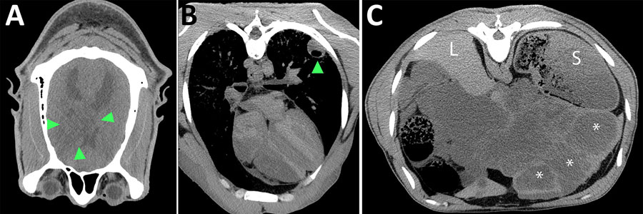
Figure 1. Postmortem computed tomography images in soft tissue window from a case of neurologic alveolar echinococcosis in postpartum zoo-housed gorilla, the Netherlands, 2024. A) Transverse view of the brain (anterior is bottom, right is left of image). Green arrows show space-occupying lesion within right hemisphere of cerebrum. B) Transverse view of the lung shows fluid- and gas-filled lesion within the left lung lobe (arrow). C) Transverse view of the liver shows multiple thick-walled, fluid-filled lesions (asterisks). L indicates normal liver tissue; S indicates stomach.
Page created: June 04, 2026
Page updated: June 26, 2026
Page reviewed: June 26, 2026
The conclusions, findings, and opinions expressed by authors contributing to this journal do not necessarily reflect the official position of the U.S. Department of Health and Human Services, the Public Health Service, the Centers for Disease Control and Prevention, or the authors' affiliated institutions. Use of trade names is for identification only and does not imply endorsement by any of the groups named above.