Volume 32, Number 7—July 2026
Research Letter
Neurologic Alveolar Echinococcosis in Postpartum Zoo-Housed Gorilla, the Netherlands, 2024
Laura A.N. Derks , Marieke Opsteegh, Denise Hoek-van Deursen, Jorrit J. Hofstra, Christine Kaandorp-Huber, Jooske IJzer, Erik A.W.S. Weerts, Volker H. Hackert, Anna R. Tellegen, Vanessa X.N. Visser, and Joke W.B. van der Giessen
Figure 2
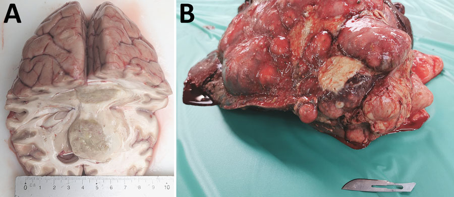
Figure 2. Necropsy brain and liver tissue from a case of neurologic alveolar echinococcosis in postpartum zoo-housed gorilla, the Netherlands, 2024. A) Cut surface of the cerebrum. Right hemisphere contains a 3.5-cm diameter, space-occupying tissue mass with ill-defined borders and secondary dislocation of preexisting structures. B) Surface of the liver. The parenchyma is largely replaced by multiple variably sized confluent nodules.
Page created: June 04, 2026
Page updated: June 26, 2026
Page reviewed: June 26, 2026
The conclusions, findings, and opinions expressed by authors contributing to this journal do not necessarily reflect the official position of the U.S. Department of Health and Human Services, the Public Health Service, the Centers for Disease Control and Prevention, or the authors' affiliated institutions. Use of trade names is for identification only and does not imply endorsement by any of the groups named above.