Volume 32, Number 7—July 2026
Research Letter
Human Pulmonary Dirofilariasis, North Queensland, Australia, 20231
Kimberley Murray, Emily Grahn, Andrew Stacey, Carly Hughes, Harsha Sheorey, Constantin Constantinoiu, Leslie Kuma, and Richard S. Bradbury
Figure 2
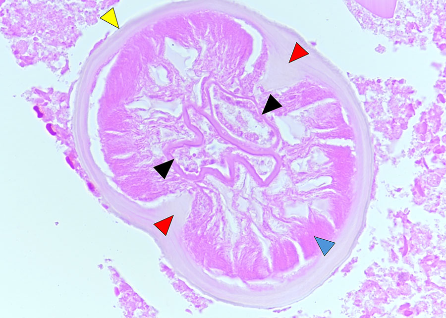
Figure 2. Defining anatomic features of Dirofilaria immitis within a small blood vessel (arteriole) in a necrotic human lung granuloma, recovered from a patient with human pulmonary dirofilariasis in Queensland, Australia, 2023. Yellow arrowhead indicates inflated necrotic smooth cuticle, without cuticular ridges; blue arrowhead indicates degenerate coelomyarian muscle structure; red arrows indicate inflated and necrotic internal cuticular ridges; and black arrows indicate degenerate paired uterine tubes. Hematoxylin and eosin stain; original magnification is ×600.
1This case was presented at the 2024 Royal College of Pathologists of Australasia Pathology Update Conference; Adelaide, Australia; 2024 Mar 1–3.
Page created: May 28, 2026
Page updated: June 26, 2026
Page reviewed: June 26, 2026
The conclusions, findings, and opinions expressed by authors contributing to this journal do not necessarily reflect the official position of the U.S. Department of Health and Human Services, the Public Health Service, the Centers for Disease Control and Prevention, or the authors' affiliated institutions. Use of trade names is for identification only and does not imply endorsement by any of the groups named above.