Volume 32, Number 8—August 2026
Dispatch
Isolation of Highly Pathogenic Avian Influenza A(H5N1) Virus from Fetal Bovine Serum, United States, 2025
Ana R. Rebelo1, Erica Butler1, Elizabeth Aguilera Nunez, Brittany Cronk, Jennifer Powers, Melissa Laverack, Chen Feng, Mohammed Nooruzzaman, Mia K. Torchetti, Leonardo C. Caserta, and Diego G. Diel
Figure 3
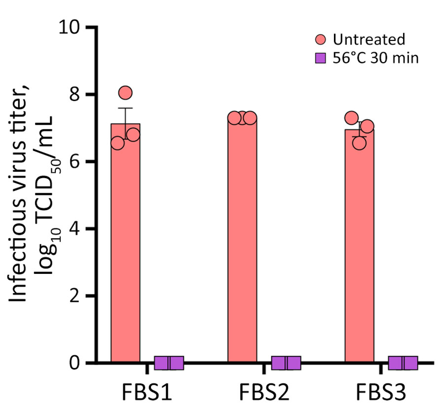
Figure 3. Heat inactivation of highly pathogenic avian influenza A virus subtype H5N1 in FBS, United States, 2025. FBS samples (1-mL aliquots) were spiked (107 TCID50/mL) with bovine highly pathogenic avian influenza A (H5N1) virus TX2/24 strain (clade 2.3.4.4b, genotype B3.13) and heat treated at 56°C for 30 minutes by using a water bath. Virus titers determined by endpoint dilutions in Cal-1 cells and expressed as TCID50/mL. Data represents observations (dots) from 3 independent experiments overlayed with the mean (horizontal line) +SEM (whiskers). FBS, fetal bovine serum; TCID50, 50% tissue culture infectious dose.
1These first authors contributed equally to this article.
Page created: June 01, 2026
Page updated: July 27, 2026
Page reviewed: July 27, 2026
The conclusions, findings, and opinions expressed by authors contributing to this journal do not necessarily reflect the official position of the U.S. Department of Health and Human Services, the Public Health Service, the Centers for Disease Control and Prevention, or the authors' affiliated institutions. Use of trade names is for identification only and does not imply endorsement by any of the groups named above.