Volume 32, Number 9—September 2026
CME ACTIVITY - Continuing Education (CE) Activity–Synopsis
Neoehrlichiosis Causing Hemolytic Anemia in Patients Treated with Rituximab, Norway, 2024–2025
 , Christine Wennerås, Hanne Quarsten, Kristine K. Berg, Anders L. Drivenes, Magnus Moksnes, and Tor Henrik A. Tvedt
Cite This Article
Citation for Media
Introduction

CDC Continuing Education (CE) ACTIVITY
PROGRAM DESCRIPTION: This activity provides the target audience with quality research to further their knowledge in neoehrlichiosis and will inform readers of the latest findings on this tickborne infection and a complication for patients taking rituximab. INTENDED AUDIENCE: Clinicians; epidemiologists; Public health departments.
Upon completion of this activity, participants will be able to:
-
Describe characteristics of neoehrlichiosis
-
Identify variables that put the three case-patients at risk for neoehrlichiosis
-
Discuss recommended treatments for neoehrlichiosis
Learning Objectives
1. Recognize the risk for public health from emerging and re-emerging infectious diseases
2. Describe characteristics of emerging infectious diseases
3. Identify populations at risk for specific emerging infectious diseases
4. Evaluate variables that put people at risk for emerging infectious diseases
5. Provide recommendations for prevention of and/or treatment for emerging and re-emerging infectious diseases
Release date: August 31, 2026; Expiration date: August 31, 2027
Authors
Martin Paulson, MD, Consultant Hematologist, Vestfold Hospital Trust; Christine Wennerås, MD, PhD, Professor, Senior Consultant, Sahlgrenska University Hospital; Hanne Quarsten, MsC, PhD, Head of the Molecular Biology Unit/Researcher, Sørlandet Hospital Trust; Kristine K. Berg, MD, Consultant MD, Sørlandet Hospital Trust, Department of Medical Microbiology; Anders L. Drivenes, MD, Resident MD, Oslo University Hospital; Magnus Moksnes, MD, Consultant MD, Vestfold Hospital Trust; Tor Henrik A. Tvedt, MD, PhD, Consultant Hematologist and Associate Professor, Oslo University Hospital, Department of Hematology.
Earning CE
To obtain CE, you should first read the journal article. After reading the article, you should be able to answer the following, related, multiple-choice questions. To complete the questions (with a minimum 80% passing score) and earn CE, please visit CDC TRAIN (https://www.train.org/cdctrain/welcome) and search for the course in the Course Catalog using SCJA5063. Follow the steps below by August 31, 2027. Credit cannot be obtained for tests completed on paper, although you may use the worksheet below to keep a record of your answers.
1. Register for and complete the course.
2. Pass the post-assessment at 80%.
All CDC continuing education courses allow two attempts to pass the post-assessment. If you do not pass on the first attempt, you have 30 days to retake it. Failing both attempts will result in a failed course status, and the course cannot be retaken during the current accreditation period. Please refer to the course description page for specific start and end dates.
3. Complete the evaluation.
4. Visit Your Learning to access your certificates and transcript.
Only one answer is correct for each question. Once you successfully answer all posttest questions and course evaluation, you will be able to view and print your certificate. No fees are charged for CDC’s CE activities.
For questions regarding this activity, contact the accredited provider, https://www.cdc.gov/cdc-train/contact. For technical assistance, contact https://www.cdc.gov/cdc-train/contact.

In support of improving patient care, the Centers for Disease Control and Prevention is jointly accredited by the Accreditation Council for Continuing Medical Education (ACCME), the Accreditation Council for Pharmacy Education (ACPE), and the American Nurses Credentialing Center (ANCC) to provide continuing education for the healthcare team.
CME: The Centers for Disease Control and Prevention designates this enduring activity for a maximum of 0.75 AMA PRA Category 1 Credits™. Physicians should claim only the credit commensurate with the extent of their participation in the activity.
AAPA CME: Credit Designation Statement—Enduring Materials: The Centers for Disease Control and Prevention has been authorized by the American Academy of PAs (AAPA) to award AAPA Category 1 CME credit for activities planned in accordance with AAPA CME Criteria. This activity is designated for 0.75 AAPA Category 1 CME credits. Approval is valid until August 31, 2029. PAs should only claim credit commensurate with the extent of their participation.
CNE: The Centers for Disease Control and Prevention designates this activity for 0.75 nursing contact hours.
CEU: The Centers for Disease Control and Prevention is authorized by IACET to offer 0.1 CEUs for this program.
AAVSB/RACE: This program was reviewed and approved by AAVSB RACE program for 1.0 hours of continuing education. Participants should be aware that some boards have limitations on the number of hours accepted in certain categories or restrictions on certain methods of delivery of continuing education. Please contact the AAVSB RACE program if you have any comments or concerns regarding this program’s validity or relevancy to the veterinary profession.
DISCLOSURE: In compliance with continuing education requirements, all planners and presenters must disclose all financial relationships, in any amount, with ineligible companies during the previous 24 months as well as any use of unlabeled product(s) or products under investigational use. CDC, our planners, and content experts wish to disclose they have no financial relationship(s) with ineligible companies with the exception of Dr. Magnus Moksnes and Dr. Tor Henrik Anderson Tvedt, and they wish to disclose the following relationships: Dr. Magnus Moksnes disclosures: Johnson & Johnson, Honoraria & advisory board; Abbvie, Honoraria; Pfizer, Honoraria; Sanofi, Advisory board; Eli Lilly, Advisory board; MDS, Advisory board; AstraZeneca, Honoraria. Dr. Tor Henrik Anderson Tvedt disclosures: Janssen, Educational material; Takeda, Advisory board/educational material. All the relevant financial relationships listed for these individuals have been mitigated. Content will not include any discussion of the unlabeled use of a product or a product under investigational use.
The Centers for Disease Control and Prevention (CDC) complies with applicable Federal civil rights laws and does not discriminate based on race, color, national origin, age, disability, religion, or sex. To learn more visit: https://www.hhs.gov/civil-rights/for-individuals/nondiscrimination/index.html.
CONTACT INFORMATION: Emerging Infectious Diseases Journal, eideditor@cdc.gov
Abstract
Neoehrlichiosis is caused by infection with the tickborne intracellular bacterium Neoehrlichia mikurensis. We describe 3 cases of direct antiglobulin test–negative hemolytic anemia in rituximab-treated patients infected with tickborne N. mikurensis bacteria in Norway during 2024–2025. All 3 patients had splenomegaly and night sweats. The hemolysis ranged from mild with compensated hemolysis to severe transfusion-dependent hemolysis that resulted in splenectomy in 1 case. All patients experienced complete resolution of hemolytic anemia and other symptoms of infection after eradication therapy with doxycycline for 21 days. Neoehrlichiosis should be considered as a curable cause of hemolytic anemia in N. mikurensis–endemic regions.
The tickborne intracellular bacterium Neoehrlichia mikurensis is the cause of the infectious disease neoehrlichiosis (1). The initial case reports of neoehrlichiosis were published from various countries in Europe in 2010 (2–4). The typical manifestations of neoehrlichiosis includes signs and symptoms of systemic inflammation, such as fever, malaise, fatigue, and nightly sweats, as well as myalgia, skin rashes and vascular events (e.g., venous thromboembolism and vasculitis) (1,5,6). Vascular manifestations probably are attributable to N. mikurensis being an intracellular pathogen that can grow and propagate in vascular endothelium (7). The infection has a variable clinical manifestation, ranging from asymptomatic carriage to life-threatening disease (3,4,8,9). The heterogenous clinical features of the disease, including manifestations such as vasculitis resembling giant cell arteritis and polyarteritis nodosa and life-threatening hemophagocytic lymphohistiocytosis, illustrate the complexity of neoehrlichiosis (6,9).
Compromised B-cell immunity and splenectomy are risk factors for neoehrlichiosis (5). Of the published cases of neoehrlichiosis, most were in patients who were immunocompromised and had frequently been treated with the B-cell lymphocyte–depleting agent rituximab (9–12). Secondary anemia is a frequent finding in patients with neoehrlichiosis that could be caused by long duration of the infection attributable to diagnostic delay (1,9,13). Neoehrlichiosis is not detected by blood culture and currently only can be diagnosed by molecular detection of bacterial DNA. One study of 103 patients with neoehrlichiosis in Sweden found a 3% prevalence of autoimmune hemolytic anemia (5). In addition, a case report describes Coombs test-positive hemolytic anemia, neutropenia, and thrombocytopenia in a dog infected with N. mikurensis (14). We describe 3 patients from southeastern Norway who had been treated with rituximab and who had splenomegaly, direct antiglobulin test (DAT)–negative hemolytic anemia, and infection with N. mikurensis.
All 3 patients were treated at Vestfold Hospital Trust, Tønsberg, Norway. Patient 1 also underwent diagnostic workup and treatment at Oslo University Hospital’s Department of Hematology (Oslo, Norway). We obtained written informed consent from all patients for the publication of their case reports.
We performed the polyspecific DAT tests for IgG and C3d at Vestfold Hospital Trust. We performed DAT tests for IgG, C3d, IgM, and IgA at Oslo University Hospital, Rikshospitalet (Oslo).
PCR Assays
We tested DNA isolated from the plasma or buffy coat fractions of whole blood by using real-time reverse transcription PCR for the detection of Borrelia burgdorferi (ospA and 16S rRNA genes), Borrelia miyamotoi (16S rRNA gene), Anaplasma phagocytophilum (groEL gene), Rickettsia spp. (gltA gene), and N. mikurensis (groEL gene, named CNM-II in the reference article) at the Department of Medical Microbiology at Sørlandet Hospital (Kristiansand, Norway) using primers and probes as described previously (8). We performed Babesia subspecies DNA PCR at Statens Serum Institut (Copenhagen, Denmark). We established a diagnosis of hemolysis on the basis of reduced hemoglobin, reduced plasma haptoglobin, increased plasma lactate dehydrogenase, and increased blood reticulocytes.
Patient 1
A 61-year-old woman sought care for rapid-onset dyspnea, fatigue, palpitations, dizziness, and a 6-month history of infrequent febrile episodes. Her medical history included rheumatoid arthritis for 17 years, breast cancer treated 16 years previously, and episodic severe depression for 1.5 years. Her arthritis initially was treated with oral methotrexate, salazopyrine, and hydroxychloroquine. Methotrexate temporarily was withdrawn because of cytopenias after a year and permanently withdrawn when she had breast cancer diagnosed. Her breast cancer was treated with surgery after 6 courses of neoadjuvant chemotherapy with fluorouracil, epirubicin hydrochloride, and cyclophosphamide, followed by postoperative radiotherapy. After completion of breast cancer treatment, treatment for rheumatoid arthritis commenced with rituximab infusions (1,000 mg 2×/y). She also was treated with venlafaxine for depression and anxiety.
At admission, we diagnosed severe transfusion-dependent direct antiglobulin test (DAT)–negative hemolytic anemia in the patient (Table). We suspected drug-induced hemolytic anemia and discontinued venlafaxine and rituximab, which later were reinstated because of lack of improvement. Two months after initially seeking care, the patient was admitted to a hospital with acute appendicitis and worsened hemolysis. After appendectomy, we initiated prednisolone (1 mg/kg/d), which resulted in partial reduction of hemolysis. We introduced the interleukin 1 receptor inhibitor anakinra 7 months later as a steroid-sparing agent after relapses of fever and worsened hemolysis during the tapering of prednisolone.
Figure
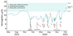
Figure. Hemoglobin levels (blue line) for patient 1 in a case series of 3 patients with neoehrlichiosis and hemolytic anemia at admission, during disease course, and after treatment with antibiotics, Norway,...
Diagnostic tests of blood and bone marrow revealed no evidence of congenital or acquired erythrocyte membrane defects, including paroxysmal nocturnal hemoglobinuria (PNH), lymphoproliferative disorders, or other hematologic neoplasia. Results of a 184-gene panel of hereditary causes of anemia and bone-marrow failure (e.g., Fanconi anemia, Diamond-Blackfan anemia, and hereditary stomatocytosis) were negative. No findings were suggestive of Wilson disease (i.e., normal ceruloplasmin and copper serum and urine levels) or of zinc- or arsenic-related poisoning. Computed tomography (CT) scans of the neck to pelvis were unremarkable, whereas a fluorodeoxyglucose (FDG) positron emission tomography (PET)–CT scan showed a slight uptake in the spleen initially deemed to be reactive. When a repeat FDG PET–CT scan 3 months later showed a maintained slight uptake in the spleen, the patient underwent diagnostic splenectomy because of a continued suspicion of underlying lymphoproliferative disorder. Before splenectomy, the patient had thrombophlebitis in the right cubital fossa and right ankle diagnosed. After splenectomy, hemoglobin levels improved for a period, but evidence of hemolysis remained. Histopathologic examination of the spleen showed depletion of B cells in the white pulp, secondary to rituximab treatment, and expanded red pulp with extramedullary hematopoiesis but no evidence of lymphoma. Within 2 months, the patient’s hemolytic anemia relapsed. Because of a history of tick exposure and erythema migrans a few years prior, we sent EDTA blood samples for PCR testing for tickborne microbes, including Rickettsia spp., B. miyamotoi, B. burgdorferi, A. phagocytophilum, N. mikurensis (8), and Babesia subsp. The patient tested positive for N. mikurensis, upon which we initiated oral doxycycline (100 mg 2×/d for 21 d). Her symptoms, most prominently fever, night sweats and fatigue, as well as all signs of hemolysis, rapidly improved and cleared completely (Table; Figure). She tested negative for N. mikurensis DNA by PCR after the course of antibiotics. We also detected N. mikurensis DNA in historical serum samples drawn 22, 11, and 6 months before the time of diagnosis. At a 12-month follow-up, no evidence of hemolytic anemia remained.
Patient 2
A 62-year-old man was referred to our outpatient clinic with a 4-month history of gradually progressive symptomatic anemia, prominent fatigue, and night sweats. He did not have fever, weight loss, nor any current or previous vascular events. His medical history included myasthenia gravis for 14 years that had been treated with rituximab infusions (1,000 mg 2×/y) for the previous 5 years. One year earlier, he had a prolonged SARS-COV-2 infection, evaluated with a chest CT, with an incidental finding of a slightly enlarged spleen. Laboratory findings at referral showed mild DAT-negative hemolytic anemia (Table). A peripheral blood smear showed no evidence of cellular dysplasia, blasts, or schistocytes. A test result for PNH was negative. We performed coagulation tests because of easy bruising, but we detected no coagulation defect. The patient had had no recall of tick bites. We performed no testing for SARS-COV-2. Because of several similarities to the first patient case (e.g., a probable nonimmunologic hemolytic anemia, treatment with rituximab, and slight splenomegaly), we performed PCR testing for tickborne microbes, including Rickettsia spp., B. miyamotoi, B. burgdorferi, A. phagocytophilum, N. mikurensis, and Babesia subsp. The patient was positive by PCR for N. mikurensis, and we initiated oral doxycycline (100 mg 2×/d for 21 d). A month later, his anemia had improved, although haptoglobin still was suppressed slightly, indicating low-grade hemolysis. Two months after the patient finished doxycycline treatment, hemoglobin and haptoglobin levels were normal (Table). At 12-month follow-up, no evidence of hemolytic anemia remained.
Patient 3
A 48-year-old man was referred our care with splenomegaly and profuse night sweats that had started 1 year earlier. His medical history included relapsing or remitting multiple sclerosis 5 years prior, which had been treated with rituximab infusions (1,000 mg 1×/y) for the previous 2 years. CT imaging of the thorax, abdomen, and pelvis done a few months before referral revealed no pathologic lymphadenopathy but substantial splenomegaly (largest diameter 17.7 cm). One month before his first episode of night sweats, he had traveled to the southern United States, Caribbean, and South America, none of which warranted malarial chemoprophylaxis. He had not contracted fever, insect bites, or rashes during his travels. He had a history of tick bites after hiking trips in southeastern Norway but no erythema migrans. Blood chemistry taken a week before the appointment at our facility showed mild and fully compensated DAT-negative hemolytic anemia (Table). The levels of haptoglobin, reticulocytes, and lactate dehydrogenase normalized spontaneously 10 days later.
An ultrasound at the time of the first consultation showed a reduction of spleen size to 15.1 cm (largest diameter). PNH test result was negative, and peripheral blood smears results were normal. Bone marrow trephine biopsy showed a very slight increase of intrasinusoidal T-lymphocytes of uncertain importance. Repeated bone marrow trephine biopsies and bone marrow aspirate analyses by flow cytometry yielded similar results. A FDG PET or CT scan showed no pathologic FDG uptake in the liver, spleen, or bone marrow and a maximal spleen diameter of 15.8 cm. We noted only a slight clonal T-cell receptor rearrangement in the bone marrow and normal results in peripheral blood, consistent with a reactive cause with no conclusive evidence of lymphoproliferative disorder. A repeat ultrasound of the spleen a few months later showed a spleen size of 14.3 cm. Six months after the initial diagnostic work-up and 2 months after receiving his yearly rituximab infusion, the patient experienced a relapse of symptoms with increasing night sweats, increase in spleen size to 17.4 cm on ultrasound, and hemolysis with a substantial reduction in hemoglobin levels and signs of intravascular hemolysis (Table).
We performed testing for tickborne microbes, including Rickettsia spp., B. miyamotoi, B. burgdorferi, A. phagocytophilum and N. mikurensis, and the patient was positive by PCR for N. mikurensis. We did not test for Babesia subsp. We initiated treatment of the patient with oral doxycycline (100 mg 2×/d for 21 d). A repeat ultrasound of the spleen performed 6 weeks later showed a reduction in size to 14 cm. His symptoms improved and resolved within a week of antibiotic treatment. A PCR test for N. mikurensis was negative after treatment. We also detected N. mikurensis DNA in stored serum samples drawn 12 and 8 months before the time of diagnosis. At 12-month follow-up, no evidence of hemolytic anemia remained.
We describe 3 patients with a diagnosis of DAT-negative hemolysis, splenomegaly, and infection with N. mikurensis. We considered neoehrlichiosis to be the cause of their hemolysis, and all patients went into remission after treatment with doxycycline. Receipt of rituximab is a risk factor for neoehrlichiosis, and splenomegaly and systemic symptoms are established features of neoehrlichiosis. Two of the 3 patients had documented N. mikurensis DNA in archival serum samples up to 2 years before the time of diagnosis. Although no archived blood samples from the second patient were available, he had splenomegaly of unknown cause 1 year previously, which in retrospect might have been caused by N. mikurensis. Neoehrlichiosis may progress from asymptomatic latent infection to symptomatic acute infection. A recent study showed that N. mikurensis causes latent infections that can reactivate when B-cell immunity is suppressed by rituximab (15). The mechanism by which N. mikurensis causes hemolysis is unclear but does not seem to involve either complement antibodies or IgG.
Hemolytic anemia invariably entails thorough review by a hematologist, especially when splenomegaly is present because lymphoproliferative disorders are a differential diagnosis. We recommend testing patients with hemolytic anemia residing in regions endemic for N. mikurensis (i.e., large parts of Europe and northern Asia) for this emerging zoonotic infection that can easily be treated and cured by oral antibiotics.
Mr. Paulson is a specialist in hematology and internal medicine at the Cancer and Hematology Center, Vestfold Hospital Trust, Tønsberg, Norway. His primary research interests include hemolytic anemia and the use of artificial intelligence in hematologic diagnostics.
References
- Grankvist A, Andersson PO, Mattsson M, Sender M, Vaht K, Höper L, et al. Infections with the tick-borne bacterium “Candidatus Neoehrlichia mikurensis” mimic noninfectious conditions in patients with B cell malignancies or autoimmune diseases. Clin Infect Dis. 2014;58:1716–22. DOIPubMedGoogle Scholar
- Welinder-Olsson C, Kjellin E, Vaht K, Jacobsson S, Wennerås C. First case of human “Candidatus Neoehrlichia mikurensis” infection in a febrile patient with chronic lymphocytic leukemia. J Clin Microbiol. 2010;48:1956–9. DOIPubMedGoogle Scholar
- von Loewenich FD, Geissdörfer W, Disqué C, Matten J, Schett G, Sakka SG, et al. Detection of “Candidatus Neoehrlichia mikurensis” in two patients with severe febrile illnesses: evidence for a European sequence variant. J Clin Microbiol. 2010;48:2630–5. DOIPubMedGoogle Scholar
- Fehr JS, Bloemberg GV, Ritter C, Hombach M, Lüscher TF, Weber R, et al. Septicemia caused by tick-borne bacterial pathogen Candidatus Neoehrlichia mikurensis. Emerg Infect Dis. 2010;16:1127–9. DOIPubMedGoogle Scholar
- Wennerås C, Wass L, Bergström B, Grankvist A, Lingblom C. Ten years of detecting Neoehrlichia mikurensis infections in Sweden: demographic, clinical and inflammatory parameters. Eur J Clin Microbiol Infect Dis. 2024;43:2083–92. DOIPubMedGoogle Scholar
- Höper L, Skoog E, Stenson M, Grankvist A, Wass L, Olsen B, et al. Vasculitis due to Candidatus Neoehrlichia mikurensis: a cohort study of 40 Swedish patients. Clin Infect Dis. 2021;73:e2372–8. DOIPubMedGoogle Scholar
- Wass L, Grankvist A, Bell-Sakyi L, Bergström M, Ulfhammer E, Lingblom C, et al. Cultivation of the causative agent of human neoehrlichiosis from clinical isolates identifies vascular endothelium as a target of infection. Emerg Microbes Infect. 2019;8:413–25. DOIPubMedGoogle Scholar
- Quarsten H, Salte T, Lorentzen ÅR, Hansen IJW, Hamre R, Forselv KJN, et al. Tick-borne pathogens detected in the blood of immunosuppressed Norwegian patients living in a tick-endemic area. Clin Infect Dis. 2021;73:e2364–71. DOIPubMedGoogle Scholar
- Gynthersen RMM, Stensvold CR, Nielsen SL, Møller HJ, Nielsen HV, Lebech AM, et al. Neoehrlichia mikurensis—an emerging opportunistic tick-borne infection in immunosuppressed patients. J Intern Med. 2023;293:782–90. DOIPubMedGoogle Scholar
- Ditlevsen Regen E, Victor D, Hendrikx T. Neoehrlichiosis in patients with multiple sclerosis in Halmstad, Sweden: a case series. J Neuroimmunol. 2026;410:
578787 . DOIPubMedGoogle Scholar - Da Silva Duarte M, Hennebel C, Fourré N, Greub G, Opota O, Merz L. Neoehrlichia mikurensis–associated secondary capillary leak syndrome and septic shock: a case report. J Med Case Rep. 2025;19:498. DOIPubMedGoogle Scholar
- Margini C, Maldonado R, Keller P, Banz Y, Escher R, Waldegg G. Fever of unknown origin, a vascular event, and immunosuppression in tick-endemic areas: think about neoehrlichiosis. Cureus. 2023;15:
e40617 . DOIPubMedGoogle Scholar - Wennerås C. Infections with the tick-borne bacterium Candidatus Neoehrlichia mikurensis. Clin Microbiol Infect. 2015;21:621–30. DOIPubMedGoogle Scholar
- Diniz PP, Schulz BS, Hartmann K, Breitschwerdt EB. “Candidatus Neoehrlichia mikurensis” infection in a dog from Germany. J Clin Microbiol. 2011;49:2059–62. DOIPubMedGoogle Scholar
- Wass L, Lewerin C, Jaén-Luchoro D, Lingblom C, Wennerås C. Latent Neoehrlichia mikurensis infections may be reactivated in patients with B-cell lymphomas treated with rituximab. Immunology. 2026;178:307–17. DOIPubMedGoogle Scholar
Figure
Table
Cite This ArticleRelated Links
Table of Contents – Volume 32, Number 9—September 2026
| EID Search Options |
|---|
 Advanced Article Search – Search articles by author and/or keyword.
|
 Articles by Country Search – Search articles by the topic country.
|
 Article Type Search – Search articles by article type and issue.
|
Please use the form below to submit correspondence to the authors or contact them at the following address:
Martin Paulson, Cancer and Hematology Center, Vestfold Hospital Trust, Halfdan Wilhelmsens allé 17, 3103 Tønsberg, Norway
Top