Volume 32, Number 10—October 2026
Synopsis
Detection of Anaplasma capra in Patients with Suspected Crimean-Congo Hemorrhagic Fever, Turkey, 2022

 , Ufuk Erol, Binnur Koksal, Omer Faruk Sahin, Ayse Nur Pektas, Seyit Ali Buyuktuna, Murtaza Oz, Yasemin Cakır Kıymaz, Tuba Nur Tasseten, Husnu Furkan Sakar, and Mehmet Bakir
Cite This Article
Citation for Media
Abstract
Anaplasma capra is a recently discovered bacterial tickborne pathogen that was first identified in goats in 2012 and later in humans in China in 2015. We detected and genetically characterized A. capra in adult patients with suspected Crimean-Congo hemorrhagic fever (CCHF) in Turkey during the 2022 outbreak. We screened all patients for A. capra by using nested PCR targeting the gltA and groEL genes. We confirmed CCHF by molecular and serological tests. Of 263 patients, 6 (2.28%) were positive for A. capra. Sequence analysis revealed 84.16%–100% gltA gene similarity and 90.24%–100% groEL gene similarity with GenBank entries. Phylogenetic analysis confirmed all A. capra gene sequences belonged to genotype 1. Our results demonstrate clinical evidence of genotype 1 A. capra infection in humans. Our findings highlight the need for clinicians to consider A. capra in the differential diagnosis of CCHF-like symptoms, especially in endemic areas.
Ticks are hematophagous ectoparasites of the subclass Acari, comprising ≈900 species worldwide. Ticks act as vectors for >200 human and animal pathogens, including bacteria such as the rickettsiae and viruses (1). The incidence and effect of tickborne diseases have risen globally because of environmental and weather changes (2). Tickborne infections impose a substantial global burden, with hundreds of thousands of human cases reported annually; Lyme disease alone is estimated to cause ≈450,000 cases each year in the United States (3,4). Turkey, situated at the crossroads of Europe and Asia, is epidemiologically critical for zoonotic infections, with 107 zoonoses reported and 21 classified as high priority by European health authorities (5,6). In Turkey, ticks serve as major vectors for various zoonotic diseases, including anaplasmosis and Crimean-Congo hemorrhagic fever (CCHF) (5–7).
Currently, CCHF is the most prevalent tickborne viral disease in humans and is endemic in parts of Africa, Asia, the Middle East, and southeastern Europe. Turkey has reported the highest number of CCHF cases globally (8–10). The first CCHF cases were documented in 2002 in the Middle Anatolia–Black Sea region, a hyperendemic area for the disease, and ≈1,000 cases have been reported annually since (10,11). The causative agent, CCHF virus (CCHFV), a member of the Nairoviridae family within the Bunyavirales order, is primarily transmitted by Hyalomma marginatum ticks. No licensed vaccine or effective antiviral treatment is available for CCHF (12). The disease typically manifests nonspecific symptoms such as fever, fatigue, myalgia, and gastrointestinal complaints but can progress to severe hemorrhagic manifestations. Case-fatality rates (CFRs) vary geographically, reaching up to 30% in some regions; in Turkey, the CFR is ≈5% (12,13).
Anaplasmosis, caused by Anaplasma marginale, A. centrale, A. phagocytophilum, A. bovis, A. ovis, and A. platys bacteria, is one of the consequential tickborne zoonotic bacterial infections and is transmitted to vertebrate hosts mainly by Ixodid species ticks. A. phagocytophilum is the etiologic agent of anaplasmosis in humans, dogs, horses, and ruminants (14). Advances in nucleic acid amplification tests and phylogenetic studies in comparing the DNA sequences of pathogens have enabled the detection of new genotypes and species of tickborne pathogens, revealing the necessity of reinterpreting the etiology of cases (14–18). In 2012, a study designed to detect Anaplasma bacteria in goats (Capra aegagrus hircus) in northern China revealed a new Anaplasma bacterial gene sequence (19). In 2015, the same gene sequence was detected in humans in China and was subsequently named A. capra. A. capra DNA was detected by nested PCR for the gltA gene in 28 (5.9%) of 477 patients who had a tick-bite history (20). Symptoms manifested as influenza-like illness, including fever, headache, malaise, dizziness, chills, gastrointestinal symptoms, and elevated hepatic enzyme concentrations. Leukopenia and thrombocytopenia were reported only in a limited number of cases (20).
Of note, ≈24% of patients with suspected CCHF in Turkey were found to be negative for CCHFV by molecular and serological tests, suggesting alternative tickborne etiologies (21). In addition, A. capra has been detected in domestic ruminants and water buffalo in the Middle Anatolia–Black Sea region of Turkey, supporting its tickborne nature and suggesting possible human infection in the same area (15,18). Our aim is to investigate the presence and molecular characteristics of A. capra in hospitalized patients with suspected CCHF in a hyperendemic region of Turkey.
Study Design and Patient Selection
This retrospective study included 263 adult patients (>18 years of age) who were hospitalized with suspected CCHF at the clinical ward of the Department of Infectious Diseases and Clinical Microbiology, Sivas Cumhuriyet University Hospital (SCUH), Sivas, Turkey, during a CCHF outbreak in 2022. SCUH is a tertiary referral hospital designated by the Turkish Ministry of Health for CCHF case management, serving a population of ≈1.5 million and located in the hyperendemic Middle Anatolia region. At admission, patients underwent a thorough physical examination and clinical history assessment by ward clinicians.
We included patients in our study on the basis of previously established case definitions (22). Study patients had >2 clinical manifestations: fever, headache, diffuse body pain, arthralgia, weakness, diarrhea, or bleeding. In addition, patients had to meet >1 criteria: residence in or recent travel to a CCHF-endemic region within the previous 2 weeks, a history of tick bite, thrombocytopenia (<1.5 × 10⁵ platelets/mm3), or leukopenia (<4 × 10³ leukocytes/mm3).
Sample Collection and CCHF Diagnosis
Three blood samples were collected from each patient. One sample was sent to the SCUH central laboratory for routine biochemical and hematological analysis; a second, 2 mL EDTA-treated (BD, https://www.bd.com) whole blood sample, was processed at the university’s Biosafety Level 2 CCHF Research Laboratory for DNA extraction; and the third sample was forwarded to the Turkish Ministry of Health’s Central Reference Virology Laboratory in Ankara for confirmation of CCHF via molecular and serologic testing.
The EDTA-treated whole blood samples were anonymized and stored in a deep freezer at −80°C) until DNA extraction. CCHF diagnosis was confirmed by a positive commercial CCHFV qualitative reverse transcriptase PCR (qRT-PCR) test kits (Bioeksen, https://www.bioeksen.com) or commercial CCHFV IgM indirect immunofluorescence test kits (Euroimmun, https://www.euroimmun.com).
We obtained epidemiologic data, including age, sex, place of residence (village, district, and city), occupation, tick exposure history, date of symptom onset, hospitalization, and sample collection via face-to-face interview records. We retrieved clinical findings, laboratory values, and outcomes (fatal or nonfatal) from the hospital’s electronic medical record system. All patients were managed by the same clinical team and received standard supportive care, including fluid resuscitation, blood product transfusions (fresh frozen plasma, packed red cells, and apheresis platelets), and cardiovascular and respiratory support as needed (22). Oral ribavirin therapy was administered at the discretion of the clinician according to a standard protocol (30 mg/kg loading dose, then 15 mg/kg/6 h for 4 d, then 7.5 mg/kg/8 h for 6 d). Patients were discharged when afebrile, without bleeding, with platelet counts >100,000/mm3, and with unremarkable coagulation parameters.
Routine microbiological testing included blood and urine cultures at admission. Complete blood count, serum biochemical tests (aspartate aminotransferase, alanine aminotransferase, creatine phosphokinase, lactate dehydrogenase), and coagulation parameters (prothrombin time and activated partial thromboplastin time) were assessed at baseline and monitored as needed during hospitalization.
Brucella spp. Bacteria Testing
In the clinical ward, testing for brucellosis is routinely performed on samples from patients with suspected CCHF (23). Diagnostic procedures include the Rose Bengal test, Wright serum agglutination test, Coombs antiglobulin test, and automated blood cultures. Clinicians interpreted the Rose Bengal test as positive or negative on the basis of visible agglutination. They performed Wright and Coombs tests by incubating patient serum at serial dilutions (>1:20) at 37°C for 48–72 hours; titers of >1:160 were considered diagnostically notable. Clinicians processed blood cultures by using BD BACTEC Plus Aerobic/F and Peds Plus/F bottles in the BD BACTEC 9120 automated system (BD). They confirmed positive cultures by using matrix-assisted laser desorption/ionization time-of-flight mass spectrometry with the MALDI Biotyper Microflex LT system (Bruker, https://www.bruker.com).
Molecular Detection of A. capra DNA
We performed nested PCR targeting the gltA gene to detect A. capra DNA in EDTA-treated blood samples. We extracted genomic DNA by using the GeneAll Exgene Clinic SV kit (GeneAll, https://geneall.com), according to the manufacturer’s protocol. In the first round of PCR, we amplified 1031 bp of the gltA gene, and in the nested PCR step, we amplified 594 bp fragments of the gltA gene by using previously described primers (Table 1). We adapted PCR protocols from previously published methods (20,24). We included positive and negative controls in each PCR run to ensure accuracy and specificity.
Genotyping of A. capra Strains
We obtained partial sequences of the gltA and groEL genes from all PCR-positive samples by using various primers (Table 1). We analyzed the nucleotide sequences by using BLAST (https://blast.ncbi.nlm.nih.gov) to determine their similarity to previously published A. capra sequences in GenBank. We submitted all the sequences generated to GenBank and obtained accession numbers for each sequence. We constructed phylogenetic trees for A. capra gltA and groEL sequences by using the maximum-likelihood method in MEGA 11 software (25). We determined the best-fit evolutionary model by using the Find Best-Fit Substitution Model tool in MEGA 11. We selected the Kimura 2-parameter model with gamma-distributed rates and invariant sites for the gltA gene (26), and we used the Tamura-Nei (TN93) model with gamma distribution among sites for the groEL gene (27). We performed bootstrap analysis with 1,000 replicates to assess tree robustness.
Data Collection and Statistical Methods
We retrospectively extracted epidemiologic, clinical, laboratory, and outcome data from the electronic medical records of SCUH and entered the data into Microsoft Excel (Microsoft, https://www.microsoft.com). Collected data included patient demographics, symptoms, physical findings, laboratory values, and outcomes (fatal vs. nonfatal). Because this was a descriptive study, we did not perform inferential statistical analyses. We have presented our results as raw counts and percentages where applicable.
Ethics Approval
The study was conducted in accordance with the Declaration of Helsinki and was approved by the research ethics committee of Sivas Cumhuriyet University (protocol no. 2022–12/16). Written informed consent was obtained from all participants.
Diagnosis of CCHF, A. capra Infection, and Brucellosis
A total of 263 patients with suspected CCHF were enrolled in the study (100 female and 163 male). Among them, 206 (78.3%) patients were confirmed to have CCHF, diagnosed via CCHFV qRT-PCR (n = 197) or CCHFV IgM serology (n = 9). Nested PCR targeting a 594-bp fragment of the A. capra gltA gene identified 6 patients (2.28%) as positive for A. capra. All 6 patients were also positive for the groEL gene. Two patients were found to have co-infections with both A. capra and CCHFV. One patient had a dual infection with A. capra and Brucella sp., which was confirmed by positive blood culture.
Among the A. capra-positive patients without co-infections, we performed an extended diagnostic workup for alternative infectious, autoimmune, and inflammatory etiologies, including viral hepatitis panels, HIV, COVID-19, Epstein-Barr virus, cytomegalovirus, Rickettsia conorii, Q fever, autoimmune antibodies (antinuclear, dsDNA, antineutrophil cytoplasmic, cyclic citrullinated peptide, rheumatoid factor, anticardiolipin), antistreptolysin O titer, and thyroid function tests. All test results were negative or unremarkable.
Demographics and Characteristics of A. capra Infection
Figure 1
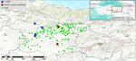
Figure 1. Geographic distribution of patients with suspected or confirmed CCHF in rural areas of Sivas, Tokat, Giresun, Yozgat, Erzincan, and Ordu Provinces, Turkey, 2022. Green dots represent confirmed CCHF patients (n...
We summarized demographic, clinical, and laboratory characteristics of the 6 patients with anaplasmosis (Table 2). Patients were 30–83 years of age; 5 were female and 1 was male. Three (50%) patients reported a history of tick exposure. All 6 were residents in rural areas of Sivas (n = 5) and Tokat (n = 1) provinces (Figure 1). Common symptoms were fatigue (100%, n = 6), musculoskeletal pain (66.7%, n = 4), headache (66.7%, n = 4), and fever history (66.7%, n = 4). Physical examination revealed splenomegaly in 2 patients (33.3%) and current fever (38.3°C) in 1 patient (16.7%). No bleeding manifestations were observed in any of the cases. Leukopenia was observed in 3 patients (50%, n = 3), including 1 co-infected with Brucella sp. (patient 4) and another with CCHFV. Thrombocytopenia was observed in 4 patients (66.7%, n = 4), 2 of whom had co-infection with CCHFV and 1 with Brucella sp. (Table 3). One patient who was co-infected with CCHFV died (Table 2).
Antimicrobial Drug Treatment for A. capra
Because of the retrospective nature of our study, patients did not receive targeted treatment for anaplasmosis during hospitalization. One surviving patient co-infected with Brucella sp. was successfully treated with a 42-day regimen of oral rifampin and doxycycline. Among the remaining patients, 1 (co-infected with CCHFV) died without receiving doxycycline therapy. The other 4 survivors were contacted and invited for postdischarge doxycycline treatment; however, they did not return for follow-up.
A. capra gltA and groEL Sequencing and Phylogenetic Analyses
We submitted the gltA and groEL gene sequences obtained from all 6 A. capra–positive patients to GenBank (accession nos. OQ819441–6 [gltA] and OQ819447–52 [groEL]). Nucleotide similarity analysis of the gltA sequences revealed 84.16%–100% identity with previously published A. capra sequences in GenBank. The sequences exhibited high similarity (98.11%–100%) to strains from cattle, sheep, goats, and wild animals across various geographic regions. In addition, our human A. capra sequences revealed 100% identity with A. capra strains previously detected in water buffalo (GenBank accession nos. ON783817–9) in Turkey, as well as with sequences from sheep (accession no. MW930534), goats (accession no. MW930535), red deer (accession no. MH084720), and swamp deer (accession no. MH084719) in France and sheep in Kyrgyzstan (accession nos. OM100820–40).
Figure 2
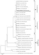
Figure 2. Phylogenetic tree of gltA gene sequences from Anaplasma capragenotype 1, genotype 2, and related species from patients with suspected or confirmed Crimean-Congo hemorrhagic fever, Turkey, 2022....
Alignment of the gltA sequences revealed 0–8 nucleotide substitutions compared with 58 A. capra sequences from domestic and wild animals. In contrast, comparisons with 158 A. capra sequences from humans, animals, and ticks showed greater genetic diversity (84.16%–88.05% similarity), with 65–67 nucleotide differences observed (Appendix Figure 1). The phylogenetic tree created on the basis of gltA sequences indicated that the sequences clustered into 2 distinct genotypes (Figure 2).
Figure 3
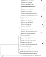
Figure 3. Phylogenetic tree of groEL gene sequences from Anaplasma capra genotype 1, genotype 2, and other Anaplasmaspecies from patients with suspected or confirmed Crimean-Congo hemorrhagic...
Similarly, the groEL gene sequences from the study samples showed 90.24%–100% identity with A. capra sequences in GenBank. We observed high nucleotide similarity (97.91%–100%) between our human A. capra sequences and those from water buffalo, sheep, goats, red deer, swamp deer, Korean water deer, roe deer, and ticks such as Dermacentor everestianus and Haemaphysalis qinghaiensis. Specifically, we noted 100% sequence identity with sequences from water buffalo in Turkey (GenBank accession nos. ON783820–2) and a sequence from a sheep in France (accession no. MW930531). Between our sequences and 26 closely related A. capra sequences, we found 0–17 nucleotide substitutions. However, we observed major differences (71–72 nucleotide substitutions) when sequences were compared with 138 A. capra sequences from domestic and wild animals, ticks, and humans (Appendix Figure 2). The phylogenetic tree we constructed from groEL sequences further supported the classification of sequences into 2 clades (Figure 3).
To date, multiple studies have demonstrated that A. capra can infect a wide range of hosts and might have a global distribution. The bacterium has been identified in various animal species, including goats, sheep (28), cattle (29), dogs (30), and yaks (31), and in multiple tick speciesm such as Haemaphysalis qinghaiensis (31), H. longicornis (32), Rhipicephalus microplus (33), Dermacentor abaensis, and D. nuttalli (34) in China. Beyond China, A. capra was first detected in cattle in Malaysia (35) and later in sheep in Sweden in 2018 (36), marking its identification in Europe. In 2021, it was also detected in cattle and ticks in Angola, establishing its presence in Africa (37). In Turkey, A. capra has been identified in cattle, sheep (15), and water buffalo (18).
Most previous studies have focused on animal hosts and the molecular characterization of A. capra. However, we detected A. capra in 6 (2.28%) of 263 adult patients who were hospitalized for suspected CCHF. This study provides clinical evidence of A. capra infection in humans outside of China, confirming its zoonotic potential and geographic spread.
Anaplasma spp. bacteria are known to infect various blood cells, including monocytes, platelets, and granulocytes, where they replicate intracellularly (14). A. capra has been shown to infect erythrocytes under in vitro conditions, forming dark vacuolar inclusions or corpuscles in the cytoplasm (38). Clinically, CCHFV infection is known to range from mild illness to severe disease with multiorgan involvement and death. In contrast, human infections with A. capra have so far been associated with milder clinical manifestations, with no reported fatalities to date. Shared clinical and laboratory features include fever, malaise, headache, myalgia, and elevated hepatic enzyme levels, whereas leukopenia and thrombocytopenia have been reported only in a limited number of A. capra cases (10,13,20). In a previous study, 5 (17.9%) of 28 patients with A. capra infection were hospitalized for 11–21 days because of severe illness characterized by lymphadenopathy and elevated liver enzymes (20). Because of the shared transmission routes and overlapping clinical features, persons living in rural areas or involved in animal husbandry are at high risk for both CCHF and anaplasmosis. The CFR for anaplasmosis is typically low (<1%); doxycycline is the drug of choice for the treatment of anaplasmosis (20,39). One study reported that all 28 patients in their cohort were successfully treated with doxycycline with no fatalities (20). In our study, 1 of the 6 patients (16.7%) died; the patient was co-infected with CCHFV.
Because CCHFV is well-established as causing death and ticks can harbor multiple pathogens (6), the specific contribution of A. capra infection to disease severity or mortality cannot be determined. However, we cannot determine whether A. capra and CCHFV and A. capra and Brucella sp. can be transmitted together by a single tick bite. This study and a previous one showed co-infections with CCHFV and other pathogens (21). Despite ticks having the potential capacity of Brucella spp. transmission, we found limited literature on tickborne brucellosis in humans (40). Because some patients are residents in rural areas and perform animal husbandry, they have risks for both CCHFV and Brucella spp. transmission.
Our findings highlight the diagnostic challenges in patients with a history of tick exposure seeking care for nonspecific clinical manifestations. Therefore, diagnostic approaches should be guided by clinical evaluation and the limitations of available laboratory tests rather than focusing on a single etiologic agent. In such settings, empiric treatment with doxycycline might be considered when clinically indicated, particularly in endemic areas for tickborne diseases.
After the recognition of A. capra’s zoonotic potential, several molecular studies have investigated its genetic characteristics. Analyses of the 16S rRNA, gltA, and groEL genes have revealed substantial genetic variation across sequences (15–18,20). Research conducted in Turkey has identified 2 distinct genotypes of A. capra, genotype 1 and genotype 2. Although prior studies found no strong correlation between genotype and host species or geographic origin, all previously reported human cases in China were associated with genotype 2 (15,17,18). In our study, both gltA and groEL gene analyses confirmed that the human A. capra sequences clustered within genotype 1 (Appendix Figures 1,2). Although genotypes and 2 can be distinguished on the basis of gene sequencing, their roles in host specificity and disease manifestations remain unclear. Further studies are needed to better characterize those genotypes and clarify their clinical and epidemiologic significance.
The first limitation of this study is that serologic testing for A. capra was not performed because of the lack of validated and widely available assays, which limits our ability to support the molecular findings with serologic evidence. Second, the small number of cases restricts the generalizability of our findings. Finally, our study population might be subject to selection bias, because our inclusion criteria required the presence of leukopenia or thrombocytopenia (because of suspected CCHF).
In conclusion, the public health significance of tickborne pathogens continues to grow because of expanding interactions between humans and tick habitats. We found molecular evidence of A. capra infection in humans in Turkey. Our findings demonstrate that A. capra is circulating not only in domestic and wild animals but also in humans in the Middle Anatolia/Black Sea region, a known hyperendemic area for CCHF. Those results confirm the zoonotic potential of A. capra genotype 1 and highlight the need for further research to identify its biologic vectors and better understand its epidemiology in CCHF-endemic areas. Clinicians working in such regions should be aware of A. capra as a differential diagnosis in patients with a history of tick exposure and suspected CCHF, especially with negative CCHFV test results.
Dr. Elaldi is a professor at the Sivas Cumhuriyet University, Sivas, Turkey. His research focuses on emerging and reemerging infections and molecular surveillance with particular emphasis on vectorborne pathogens, including Crimean-Congo hemorrhagic fever virus.
Acknowledgments
The authors gratefully acknowledge the Central CCHF Reference Virology Laboratory of the Turkish Ministry of Health, Ankara, Turkey, for providing diagnostic testing for CCHFV.
Author contributions: conceptualization, methodology, formal analysis, original draft, and review and editing of draft, K.A. and N.E.; methodology and formal analysis, U.E., B.K., O.F.S., and A.N.P.; methodology and resources, S.A.B., M.O., Y.C.K., T.N.T., H.F.S., and M.B.
References
- Negi T, Kandari LS, Arunachalam K. Update on prevalence and distribution pattern of tick-borne diseases among humans in India: a review. Parasitol Res. 2021;120:1523–39. DOIPubMedGoogle Scholar
- Sonenshine DE. Range expansion of tick disease vectors in North America: implications for spread of tick-borne disease. Int J Environ Res Public Health. 2018;15:478. DOIPubMedGoogle Scholar
- de la Fuente J, Estrada-Pena A, Venzal JM, Kocan KM, Sonenshine DE. Overview: ticks as vectors of pathogens that cause disease in humans and animals. Front Biosci. 2008;13:6938–46. DOIPubMedGoogle Scholar
- Centers for Disease Control and Prevention. Lyme disease surveillance and data, 2025 [cited 2026 Mar 23]. https://www.cdc.gov/lyme/data-research/facts-stats/index.html
- Düzlü Ö, İnci A, Yıldırım A, Doğanay M, Özbel Y, Aksoy S. Vector-borne zoonotic diseases in Turkey: rising threats on public health. Turkiye Parazitol Derg. 2020;44:168–75. DOIPubMedGoogle Scholar
- Inci A, Yildirim A, Duzlu O, Doganay M, Aksoy S. Tick-borne diseases in Turkey: a review based on one health perspective. PLoS Negl Trop Dis. 2016;10:
e0005021 . DOIPubMedGoogle Scholar - Aydin L, Bakirci S. Geographical distribution of ticks in Turkey. Parasitol Res. 2007;101(Suppl 2):S163–6. DOIPubMedGoogle Scholar
- Sorvillo TE, Rodriguez SE, Hudson P, Carey M, Rodriguez LL, Spiropoulou CF, et al. Towards a sustainable One Health approach to Crimean-Congo hemorrhagic fever prevention: focus areas and gaps in knowledge. Trop Med Infect Dis. 2020;5:113. DOIPubMedGoogle Scholar
- Shahhosseini N, Wong G, Babuadze G, Camp JV, Ergonul O, Kobinger GP, et al. Crimean-Congo hemorrhagic fever virus in Asia, Africa and Europe. Microorganisms. 2021;9:1907. DOIPubMedGoogle Scholar
- Bente DA, Forrester NL, Watts DM, McAuley AJ, Whitehouse CA, Bray M. Crimean-Congo hemorrhagic fever: history, epidemiology, pathogenesis, clinical syndrome and genetic diversity. Antiviral Res. 2013;100:159–89. DOIPubMedGoogle Scholar
- Gunes T, Engin A, Poyraz O, Elaldi N, Kaya S, Dokmetas I, et al. Crimean-Congo hemorrhagic fever virus in high-risk population, Turkey. Emerg Infect Dis. 2009;15:461–4. DOIPubMedGoogle Scholar
- Hawman DW, Feldmann H. Crimean-Congo haemorrhagic fever virus. Nat Rev Microbiol. 2023;21:463–77. DOIPubMedGoogle Scholar
- Yilmaz GR, Buzgan T, Irmak H, Safran A, Uzun R, Cevik MA, et al. The epidemiology of Crimean-Congo hemorrhagic fever in Turkey, 2002–2007. Int J Infect Dis. 2009;13:380–6. DOIPubMedGoogle Scholar
- Dumler JS, Barbet AF, Bekker CP, Dasch GA, Palmer GH, Ray SC, et al. Reorganization of genera in the families Rickettsiaceae and Anaplasmataceae in the order Rickettsiales: unification of some species of Ehrlichia with Anaplasma, Cowdria with Ehrlichia and Ehrlichia with Neorickettsia, descriptions of six new species combinations and designation of Ehrlichia equi and ‘HGE agent’ as subjective synonyms of Ehrlichia phagocytophila. Int J Syst Evol Microbiol. 2001;51:2145–65. DOIPubMedGoogle Scholar
- Altay K, Erol U, Sahin OF. The first molecular detection of Anaplasma capra in domestic ruminants in the central part of Turkey, with genetic diversity and genotyping of Anaplasma capra. Trop Anim Health Prod. 2022;54:129. DOIPubMedGoogle Scholar
- Altay K, Erol U, Sahin OF, Aytmirzakizi A. First molecular detection of Anaplasma species in cattle from Kyrgyzstan; molecular identification of human pathogenic novel genotype Anaplasma capra and Anaplasma phagocytophilum related strain. Ticks Tick Borne Dis. 2022;13:
101861 . DOIPubMedGoogle Scholar - Altay K, Erol U, Sahin OF, Aytmirzakizi A, Temizel EM, Aydin MF, et al. The detection and phylogenetic analysis of Anaplasma phagocytophilum-like 1, A. ovis and A. capra in sheep: A. capra divides into two genogroups. Vet Res Commun. 2022;46:1271–9. DOIPubMedGoogle Scholar
- Sahin OF, Erol U, Altay K. Buffaloes as new hosts for Anaplasma capra: Molecular prevalence and phylogeny based on gtlA, groEL, and 16S rRNA genes. Res Vet Sci. 2022;152:458–64. DOIPubMedGoogle Scholar
- Liu Z, Ma M, Wang Z, Wang J, Peng Y, Li Y, et al. Molecular survey and genetic identification of Anaplasma species in goats from central and southern China. Appl Environ Microbiol. 2012;78:464–70. DOIPubMedGoogle Scholar
- Li H, Zheng YC, Ma L, Jia N, Jiang BG, Jiang RR, et al. Human infection with a novel tick-borne Anaplasma species in China: a surveillance study. Lancet Infect Dis. 2015;15:663–70. DOIPubMedGoogle Scholar
- Fırtına Topcu K, Sümer Z. Investigation of the Crimean-Congo hemorrhagic fever virus and the seropositivity of Coxiella burnetii, Borrelia burgdorferi in patients with a history of tick bites. Turk Mikrobiyol Cemiy Derg. 2022;52:265–73. DOIGoogle Scholar
- Leblebicioglu H, Bodur H, Dokuzoguz B, Elaldi N, Guner R, Koksal I, et al. Case management and supportive treatment for patients with Crimean-Congo hemorrhagic fever. Vector Borne Zoonotic Dis. 2012;12:805–11. DOIPubMedGoogle Scholar
- Öz M, Çubuk F, Çakır Kıymaz Y, Öksüz C, Hasbek M, Büyüktuna SA, et al. Hidden threats: brucellosis diagnosis and co-infection patterns in Crimean-Congo hemorrhagic fever suspects. Diagn Microbiol Infect Dis. 2025;111:
116724 . DOIPubMedGoogle Scholar - Yang J, Liu Z, Niu Q, Liu J, Han R, Liu G, et al. Molecular survey and characterization of a novel Anaplasma species closely related to Anaplasma capra in ticks, northwestern China. Parasit Vectors. 2016;9:603. DOIPubMedGoogle Scholar
- Tamura K, Stecher G, Kumar S. MEGA11: molecular evolutionary genetics analysis version 11. Mol Biol Evol. 2021;38:3022–7. DOIPubMedGoogle Scholar
- Kimura M. A simple method for estimating evolutionary rates of base substitutions through comparative studies of nucleotide sequences. J Mol Evol. 1980;16:111–20. DOIPubMedGoogle Scholar
- Tamura K, Nei M. Estimation of the number of nucleotide substitutions in the control region of mitochondrial DNA in humans and chimpanzees. Mol Biol Evol. 1993;10:512–26.PubMedGoogle Scholar
- Yang J, Liu Z, Niu Q, Liu J, Han R, Guan G, et al. A novel zoonotic Anaplasma species is prevalent in small ruminants: potential public health implications. Parasit Vectors. 2017;10:264. DOIPubMedGoogle Scholar
- Guo WP, Huang B, Zhao Q, Xu G, Liu B, Wang YH, et al. Human-pathogenic Anaplasma spp., and Rickettsia spp. in animals in Xi’an, China. PLoS Negl Trop Dis. 2018;12:
e0006916 . DOIPubMedGoogle Scholar - Shi K, Li J, Yan Y, Chen Q, Wang K, Zhou Y, et al. Dogs as new hosts for the emerging zoonotic pathogen Anaplasma capra in China. Front Cell Infect Microbiol. 2019;9:394. DOIPubMedGoogle Scholar
- Wang Y, Zhang Q, Han S, Li Y, Wang B, Yuan G, et al. Ehrlichia chaffeensis and four Anaplasma species with veterinary and public health significance identified in Tibetan sheep (Ovis aries) and yaks (Bos grunniens) in Qinghai, China. Front Vet Sci. 2021;8:
727166 . DOIPubMedGoogle Scholar - Zhang H, Sun Y, Jiang H, Huo X. Prevalence of severe febrile and thrombocytopenic syndrome virus, Anaplasma spp. and Babesia microti in hard ticks (Acari: Ixodidae) from Jiaodong Peninsula, Shandong Province. Vector Borne Zoonotic Dis. 2017;17:134–40. DOIPubMedGoogle Scholar
- Guo WP, Zhang B, Wang YH, Xu G, Wang X, Ni X, et al. Molecular identification and characterization of Anaplasma capra and Anaplasma platys-like in Rhipicephalus microplus in Ankang, Northwest China. BMC Infect Dis. 2019;19:434. DOIPubMedGoogle Scholar
- Han R, Yang JF, Mukhtar MU, Chen Z, Niu QL, Lin YQ, et al. Molecular detection of Anaplasma infections in ixodid ticks from the Qinghai-Tibet Plateau. Infect Dis Poverty. 2019;8:12. DOIPubMedGoogle Scholar
- Koh FX, Panchadcharam C, Sitam FT, Tay ST. Molecular investigation of Anaplasma spp. in domestic and wildlife animals in Peninsular Malaysia. Vet Parasitol Reg Stud Reports. 2018;13:141–7. DOIPubMedGoogle Scholar
- Grandi G, Aspán A, Pihl J, Gustafsson K, Engström F, Jinnerot T, et al. Detection of tick-borne pathogens in lambs undergoing prophylactic treatment against ticks on two Swedish farms. Front Vet Sci. 2018;5:72. DOIPubMedGoogle Scholar
- Barradas PF, Mesquita JR, Ferreira P, Gärtner F, Carvalho M, Inácio E, et al. Molecular identification and characterization of Rickettsia spp. and other tick-borne pathogens in cattle and their ticks from Huambo, Angola. Ticks Tick Borne Dis. 2021;12:
101583 . DOIPubMedGoogle Scholar - Peng Y, Lu C, Yan Y, Song J, Pei Z, Gong P, et al. The novel zoonotic pathogen, Anaplasma capra, infects human erythrocytes, HL-60, and TF-1 cells in vitro. Pathogens. 2021;10:600. DOIPubMedGoogle Scholar
- MacQueen D, Centellas F. Human granulocytic anaplasmosis. Infect Dis Clin North Am. 2022;36:639–54. DOIPubMedGoogle Scholar
- Ma R, Li C, Gao A, Jiang N, Feng X, Li J, et al. Evidence-practice gap analysis in the role of tick in brucellosis transmission: a scoping review. Infect Dis Poverty. 2024;13:3. DOIPubMedGoogle Scholar
Figures
Tables
Cite This ArticleOriginal Publication Date: September 16, 2026
1These first authors contributed equally to this article.
Table of Contents – Volume 32, Number 10—October 2026
| EID Search Options |
|---|
 Advanced Article Search – Search articles by author and/or keyword.
|
 Articles by Country Search – Search articles by the topic country.
|
 Article Type Search – Search articles by article type and issue.
|
Please use the form below to submit correspondence to the authors or contact them at the following address:
Nazif Elaldi, Sivas Cumhuriyet University Faculty of Medicine, Department of Infectious Diseases and Clinical Microbiology, 58140, Sivas, Turkey
Top