Skip directly to site content Skip directly to page options Skip directly to A-Z link Skip directly to A-Z link Skip directly to A-Z link
Volume 5, Number 6—December 1999
Perspective

Emerging Infectious Diseases and Amphibian Population Declines

Peter Daszak*Comments to Author , Lee Berger†‡, Andrew A. Cunningham§, Alex D. Hyatt†, D. Earl Green¶, and Rick Speare‡
Author affiliations: *Institute of Ecology, University of Georgia, Athens, Georgia, USA; †Australian Animal Health Laboratory, Commonwealth Scientific Industrial Research Organization, Geelong, Victoria, Australia; ‡School of Public Health and Tropical Medicine, James Cook University, Townsville, Queensland, Australia; §Institute of Zoology, Zoological Society of London, London, United Kingdom; and ¶National Wildlife Health Center, U.S. Geological Survey, Madison, Wisconsin, USA

Main Article

Figure 1

. A: Ventral abdominal skin of Bufo haematiticus from western Panama. The superficial keratinized layer of epidermis (stratum corneum) contains numerous intracellular spherical-to-ovoid sporangia (spore-containing bodies) of Batrachochytrium sp. The mature sporangia (sp, arrows) are 12-20 µm (n = 25) in diameter and have refractile walls 0.5-2.0 µm thick. Most sporangia are empty, having discharged all zoospores, but a few sporangia contain two to nine zoospores. This stratum corneum is markedly

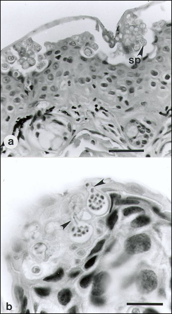
Figure 1. . A: Ventral abdominal skin of Bufo haematiticus from western Panama. The superficial keratinized layer of epidermis (stratum corneum) contains numerous intracellular spherical-to-ovoid sporangia (spore-containing bodies) of Batrachochytrium sp. The mature sporangia (sp, arrows) are 12-20 µm (n = 25) in diameter and have refractile walls 0.5-2.0 µm thick. Most sporangia are empty, having discharged all zoospores, but a few sporangia contain two to nine zoospores. This stratum corneum is markedly thickened adjacent to groups of parasitized cells and in some cases, the superficial layer has become detached. No chytrids are present in the stratum spinosum, stratum basale, dermis, dermal glands, and blood vessels. Note the absence of hyphae and lack of an inflammatory cell response in the deeper layers of epidermis and the dermis. Hematoxylin and eosin stain. Bar = 35 µm. 1b. Ventral skin of upper hind limb of Atelopus varius from western Panama. Two sporangia containing numerous zoospores are visible within cells of the stratum corneum. Each flask-shaped sporangium has a single characteristic discharge tube (arrow) at the skin surface. Exiting zoospores are visible in the discharge tubes of both sporangia. Hyperkeratosis is minimal in this acute infection. Tissues were fixed in neutral-buffered 10% formalin, paraffin-embedded, sectioned at 6 µm thick and stained with hematoxylin and eosin. Bar = 35 µm.

Main Article

1Note that the relative number of mass death events decreases with increasing impact on population.

Page created: December 15, 2010
Page updated: December 15, 2010
Page reviewed: December 15, 2010
The conclusions, findings, and opinions expressed by authors contributing to this journal do not necessarily reflect the official position of the U.S. Department of Health and Human Services, the Public Health Service, the Centers for Disease Control and Prevention, or the authors' affiliated institutions. Use of trade names is for identification only and does not imply endorsement by any of the groups named above.
file_external