Volume 6, Number 3—June 2000
Research
Rhinosporidium seeberi: A Human Pathogen from a Novel Group of Aquatic Protistan Parasites
Figure 4
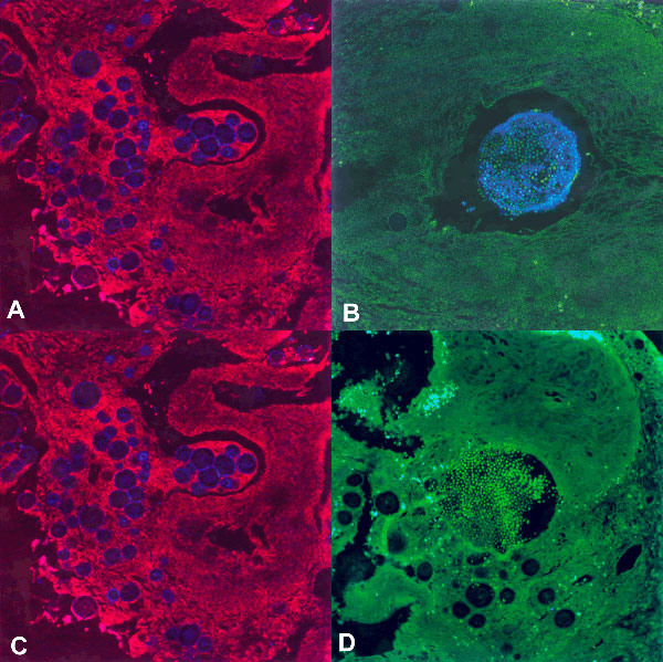
Figure 4. An oligonucleotide probe complementary to a unique region of the Rhinosporidium seeberi 18S rRNA sequence localizes to visible organisms in human nasal tissue by using fluorescence in situ hybridization. Confocal micrographs at 200X magnification. The Cy-5-labeled Rhinosporidium probe (blue) hybridizes to spherical R. seeberi trophocytes (A) and a sporangium with endospores (B). A control probe consisting of the complement of the Rhinosporidium probe labeled with Cy-5 does not hybridize to the trophocytes (C) or a sporangium with endospores (D). Images were collected in two wave-length channels: the first for Texas Red (red) or FITC (green) displays tissue architecture through autofluorescence, and the second channel for Cy-5 (blue pseudocolor) displays the probe signal.