Volume 10, Number 11—November 2004
Research
Topographic Changes in SARS Coronavirus–infected Cells during Late Stages of Infection
Mah-Lee Ng* , J.W.M. Lee*, M.L.N. Leong*, A.-E. Ling†, H.-C. Tan‡, and E.E. Ooi‡
Figure 1
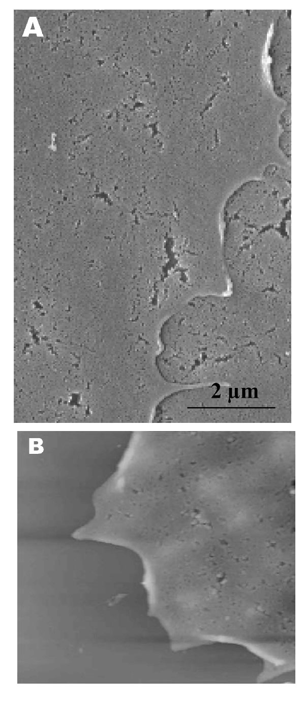
Figure 1. Scanning electron (A) and atomic force (B) microscopy images of uninfected Vero cells. A) Under the scanning electron microscope, uninfected cells look relatively flat with minimal surface morphology. No pronounced pseudopodia are visible on the cell edge or surfaces. B) Atomic force microscopy confirms the form and structure seen in panel A. Cell surface is uniformly flat.
Page created: April 21, 2011
Page updated: April 21, 2011
Page reviewed: April 21, 2011
The conclusions, findings, and opinions expressed by authors contributing to this journal do not necessarily reflect the official position of the U.S. Department of Health and Human Services, the Public Health Service, the Centers for Disease Control and Prevention, or the authors' affiliated institutions. Use of trade names is for identification only and does not imply endorsement by any of the groups named above.