Volume 10, Number 11—November 2004
Research
Topographic Changes in SARS Coronavirus–infected Cells during Late Stages of Infection
Mah-Lee Ng* , J.W.M. Lee*, M.L.N. Leong*, A.-E. Ling†, H.-C. Tan‡, and E.E. Ooi‡
Figure 7
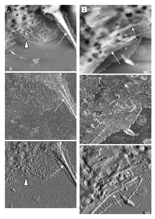
Figure 7. Vero cells infected with severe acute respiratory syndrome–associated coronavirus at 15 h after infection. A and B) When the hard tapping mode of the atomic force microscope is used, thickened cell cytoskeletal filaments are found below the subcellular surface of the cells. A) Enhanced cytoskeletal network of the cytoplasmic region (arrowhead). Arrow shows the much-thickened filaments within the pseudopodia of the cell. B) At high resolution, the arrows show the thickened cytoskeletal filaments along the cell periphery. The height and amplitude images clearly show the cytoskeletal filaments parallel to the cell edge.
Page created: April 21, 2011
Page updated: April 21, 2011
Page reviewed: April 21, 2011
The conclusions, findings, and opinions expressed by authors contributing to this journal do not necessarily reflect the official position of the U.S. Department of Health and Human Services, the Public Health Service, the Centers for Disease Control and Prevention, or the authors' affiliated institutions. Use of trade names is for identification only and does not imply endorsement by any of the groups named above.