Skip directly to site content Skip directly to page options Skip directly to A-Z link Skip directly to A-Z link Skip directly to A-Z link
Volume 10, Number 3—March 2004
Research

Monkeypox Transmission and Pathogenesis in Prairie Dogs

Jeannette Guarner*Comments to Author , Bill J. Johnson†, Christopher D. Paddock*, Wun-Ju Shieh*, Cynthia S. Goldsmith*, Mary G. Reynolds*, Inger K. Damon*, Russell L. Regnery*, Sherif R. Zaki*, and the Veterinary Monkeypox Virus Working Group
Author affiliations: *Centers for Disease Control and Prevention, Atlanta, Georgia, USA; †Oklahoma Animal Disease Diagnostic Laboratory, Oklahoma State University, Stillwater, Oklahoma, USA

Main Article

Figure 2

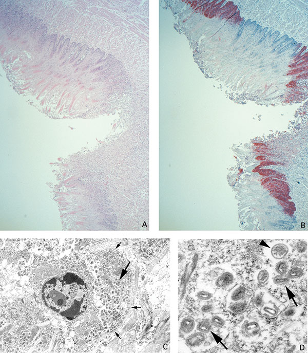
Ulcer on tongue of a prairie dog infected with monkeypox virus (A: hematoxylin and eosin stain, 12.5X original magnification). Orthopox viral antigens are abundant in the squamous epithelium, with lesser amounts in the ulcer bed (B: immunohistochemical stain using the anti-smallpox antibody, 12.5X original magnification). Tongue epithelial cell adjacent to epidermal basement membrane (small arrows) with Guarnieri-like inclusion (large arrow) (C: transmission electron microscopy, 2,400X original

Figure 2. Ulcer on tongue of a prairie dog infected with monkeypox virus (A: hematoxylin and eosin stain, 12.5X original magnification). Orthopox viral antigens are abundant in the squamous epithelium, with lesser amounts in the ulcer bed (B: immunohistochemical stain using the anti-smallpox antibody, 12.5X original magnification). Tongue epithelial cell adjacent to epidermal basement membrane (small arrows) with Guarnieri-like inclusion (large arrow) (C: transmission electron microscopy, 2,400X original magnification). Higher magnification of the Guarnieri-like inclusion shows intracellular immature (arrowhead) and mature (arrows) orthopox virions. The mature virions consist of a dense core surrounded by several laminated zones and enclosed within an outer membrane. (D: transmission electron microscopy, 17,000X original magnification.)

Main Article

1Veterinary Monkeypox Virus Working Group Members: CDC’s Infectious Diseases Pathology Activity: Patricia Greer, Michelle M. Packard, and William Lee, and CDC’s Poxvirus Section Laboratory: Victoria Olson, Richard Kline, Yu Li, and Linda Stempora.

Page created: February 08, 2011
Page updated: February 08, 2011
Page reviewed: February 08, 2011
The conclusions, findings, and opinions expressed by authors contributing to this journal do not necessarily reflect the official position of the U.S. Department of Health and Human Services, the Public Health Service, the Centers for Disease Control and Prevention, or the authors' affiliated institutions. Use of trade names is for identification only and does not imply endorsement by any of the groups named above.
file_external