Volume 11, Number 4—April 2005
Research
Experimental Infection of Prairie Dogs with Monkeypox Virus
Figure 2
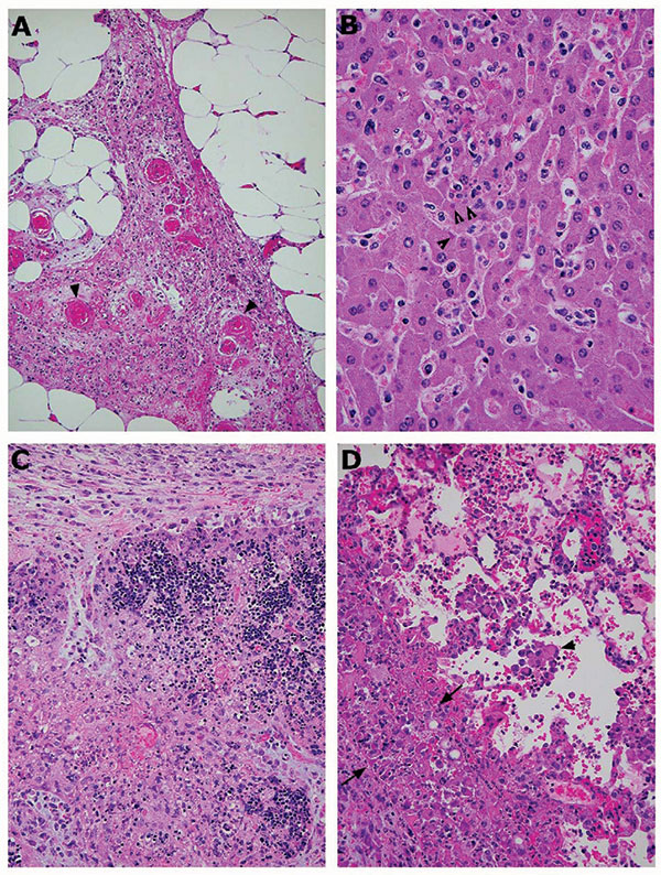
Figure 2. Photomicrographs of pathologic changes in tissues of prairie dogs infected with monkeypox virus. A) Abdominal adipose tissue from an intraperitoneally infected animal showing focal vasculitis (arrowheads), necrosis, and proliferation of fibroblasts. B) Mild hepatitis, characterized by focal inflammatory cell infiltration in the lobules and hepatocytes containing cytoplasmic inclusion bodies (arrowheads). C) Severe necrosis of the thymus from an animal infected intranasally. The necrotic areas contain many swollen macrophages with cytoplasm full of intensively eosinophilic material (viral proteins). D) Lung from the same animal, showing inflammation with swollen cells (arrowheads), alveolar edema, and necrosis of the pleura. The latter is infiltrated by many inclusion-filled macrophages and proliferating fibroblasts (the thick layer between the 2 arrows). Magnification: A, 10× objective; B, 40× objective; C, 10× objective; D, 20× objective. Hematoxylin and eosin stain.