Volume 13, Number 12—December 2007
Research
Susceptibility of Canada Geese (Branta canadensis) to Highly Pathogenic Avian Influenza Virus (H5N1)
John Pasick* , Yohannes Berhane*, Carissa Embury-Hyatt*, John Copps*, Helen Kehler*, Katherine Handel*, Shawn Babiuk*, Kathleen Hooper-McGrevy*, Yan Li†, Quynh Mai Le‡, and Song Lien Phuong§
Figure 1
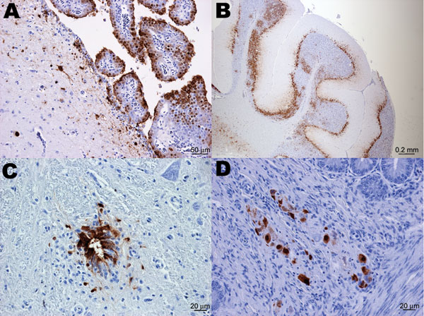
Figure 1. Immunohistochemical staining for influenza virus nucleoprotein in central and peripheral nervous system of naive juvenile Canada geese tissues after challenge with influenza virus (H5N1). A) Cerebrum. Positive immunolabeling of neurons, glial cells, ependymal and choroid plexus epithelial cells. B) Cerebellum. Extensive positive immunolabeling of Purkinje cells and neurons of the granular layer. C) Spinal cord. Positive immunolabeling of ependymal cells of the central canal and adjacent neurons and glial cells. D) Small intestine. Positive immunolabeling of neurons of the submucosal plexus.
Page created: July 06, 2010
Page updated: July 06, 2010
Page reviewed: July 06, 2010
The conclusions, findings, and opinions expressed by authors contributing to this journal do not necessarily reflect the official position of the U.S. Department of Health and Human Services, the Public Health Service, the Centers for Disease Control and Prevention, or the authors' affiliated institutions. Use of trade names is for identification only and does not imply endorsement by any of the groups named above.