Volume 13, Number 12—December 2007
Research
Phenotypic Similarity of Transmissible Mink Encephalopathy in Cattle and L-type Bovine Spongiform Encephalopathy in a Mouse Model
Figure 4
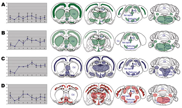
Figure 4. Brain lesion profiles (left panels) and disease-associated prion protein brain distribution (right panels) observed in the brain of TgOvPrP4 mice infected with L-type bovine spongiform encephalopathy (BSE), at first (A, n = 5) and second (B, n = 4) passages; TME in cattle (C, n = 5); or typical BSE (D, n = 4) at second passage. Brain vacuolation was scored (means ± standard deviations) on a scale of 0–5 in the following brain areas: 1) dorsal medulla nuclei, 2) cerebellar cortex, 3) superior colliculus, 4) hypothalamus, 5) central thalamus, 6) hippocampus, 7) lateral septal nuclei, 8) cerebral cortex at the level of thalamus, and 9) cerebral cortex at the level of septal nuclei. In right panels, showing the PrPd distribution, stars indicate the presence of florid plaques.