Volume 13, Number 3—March 2007
Research
In Vitro Cell Culture Infectivity Assay for Human Noroviruses
Timothy M. Straub* , Kerstin Höner zu Bentrup†, Patricia Orosz Coghlan‡, Alice Dohnalkova*, Brooke K. Mayer*1, Rachel A. Bartholomew*, Catherine O. Valdez*, Cynthia J. Bruckner-Lea*, Charles P. Gerba‡, Morteza A. Abbaszadegan§, and Cheryl A. Nickerson†1
Figure 3
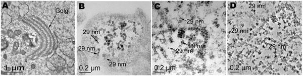
Figure 3. Transmission electron microscopy of uninfected and infected cell cultures from the second infection trial. A) Uninfected cells showing normal internal membrane organelles. B) Suspect 29-nm particles in cells, viruses from cell culture lysate from the first infection trial (P1). C) Stool sample flag2 (P0) and D) stool sample 149 (P0) showing numerous 29-nm particles and internal rearrangement of membrane-bound organelles.
Page created: June 29, 2010
Page updated: June 29, 2010
Page reviewed: June 29, 2010
The conclusions, findings, and opinions expressed by authors contributing to this journal do not necessarily reflect the official position of the U.S. Department of Health and Human Services, the Public Health Service, the Centers for Disease Control and Prevention, or the authors' affiliated institutions. Use of trade names is for identification only and does not imply endorsement by any of the groups named above.