Volume 5, Number 6—December 1999
Perspective
Emerging Infectious Diseases and Amphibian Population Declines
Peter Daszak* , Lee Berger†‡, Andrew A. Cunningham§, Alex D. Hyatt†, D. Earl Green¶, and Rick Speare‡
Figure 4
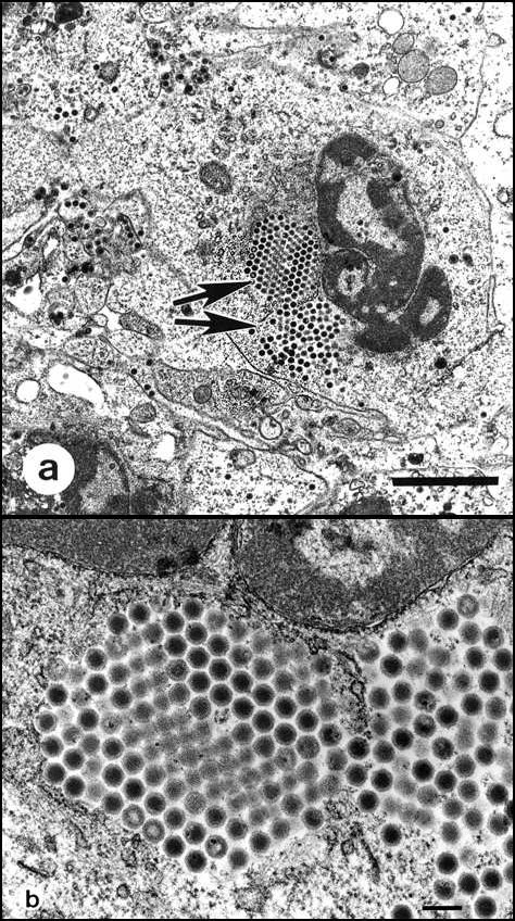
Figure 4. Transmission electron micrographs of iridovirus cultured from the liver of a naturally diseased common frog (Rana temporaria) by using a fathead minnow epithelial cell line. 4a. Virus-infected cell. Large isocahedral viruses are conspicuous within the cytoplasm (arrows). Bar = 2 µm. 4b. Paracrystalline array of iridovirus. Bar = 200 µm.
Page created: December 15, 2010
Page updated: December 15, 2010
Page reviewed: December 15, 2010
The conclusions, findings, and opinions expressed by authors contributing to this journal do not necessarily reflect the official position of the U.S. Department of Health and Human Services, the Public Health Service, the Centers for Disease Control and Prevention, or the authors' affiliated institutions. Use of trade names is for identification only and does not imply endorsement by any of the groups named above.