Volume 10, Number 9—September 2004
Research
Experimental Infection of Ground Squirrels (Spermophilus tridecemlineatus) with Monkeypox Virus
Figure 1
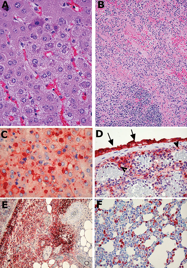
Figure 1. Representative photomicrographs of histologic changes and immunohistochemical staining of tissues from ground squirrels infected with monkeypox virus. A) Liver from a ground squirrel (intranasal infection) showing mild degenerative changes, including early steatosis, and purple-colored viral cytoplasmic inclusion bodies in the hepatocytes (40x objective). B) Spleen from a ground squirrel infected intraperitoneally, showing extensive necrosis (20x objective). C) Liver showing positive antigen staining of the intrahepatocytic inclusion bodies; antigen is present in the cytoplasm and to a lesser extent in cell membranes (40x objective). D) Spleen from a ground squirrel infected intraperitoneally, showing positive antigen staining in the interstitial cells, endothelial cells (arrowheads), and the surface mesothelial cells (arrows) (20x objective). E) Same tissue sample as D, showing the edge of the spleen with antigen-positive mesothelial layer; the adjacent fat and fibrous tissue show necrosis but are also strongly positive for antigen (20x objective). F) Lung from the same animal showing many antigen-positive interstitial cells and pneumocytes (40x objective). A and B, hematoxylin and eosin stain; C–F, immunoperoxidase stain.