Volume 10, Number 9—September 2004
Research
Experimental Infection of Ground Squirrels (Spermophilus tridecemlineatus) with Monkeypox Virus
Robert B. Tesh* , Douglas M. Watts*, Elena Sbrana*, Marina Siirin*, Vsevolod L. Popov*, and Shu-Yuan Xiao*
Figure 2
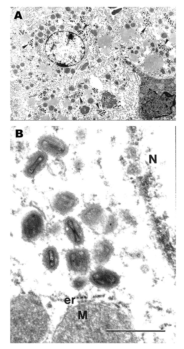
Figure 2. Ultrastructural localization of monkeypox virus in hepatocytes in the liver of a ground squirrel 5 days after infection. A) Hepatocytes contain numerous groups of virions (arrows) in their cytoplasm (bar = 1 μm). B) Magnified area of A, showing typical ultrastructure of monkeypox virus virions and characteristic hepatocyte mitochondria (M) surrounded by cisterns of granular endoplasmic reticulum (er). N, fragment of hepatocyte nucleus; bar = 0.5 μm.
Page created: March 30, 2011
Page updated: March 30, 2011
Page reviewed: March 30, 2011
The conclusions, findings, and opinions expressed by authors contributing to this journal do not necessarily reflect the official position of the U.S. Department of Health and Human Services, the Public Health Service, the Centers for Disease Control and Prevention, or the authors' affiliated institutions. Use of trade names is for identification only and does not imply endorsement by any of the groups named above.