Volume 12, Number 6—June 2006
Research
Co-infections of Adenovirus Species in Previously Vaccinated Patients
Figure
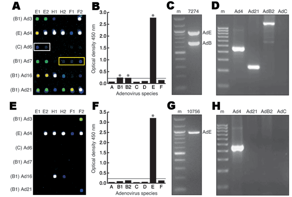
Figure. Molecular methods used to identify human adenovirus (HAdV) co-infections. A–D) Vaccinated sample 7274. A) Microarray hybridization profile. White and yellow rectangles indicate low-positive HAdV-C and HAdV-B2 targets, respectively. Spot colors denote hybridization signal intensity (white > yellow > green > blue). Species and corresponding serotype designations are indicated on the left. Probe designations (E1, E2 = serotype-specific E1A probes; H1, H2 = serotype-specific hexon probes; F1, F2 = serotype-specific fiber probes) are indicated above each array. B) Adenovirus Consensus kit optical density values. *, amplification positive. The horizontal line is the manufacturer's significance threshhold. C) Multiplex species-specific polymerase chain reaction (PCR). m, molecular mass marker. Species designations are to the right of the corresponding band. D) PCR verification with independent serotype or species-specific primers. E–H) Unvaccinated sample 10756. E) Microarray hybridization profile. F) Adenovirus Consensus kit optical density values. G) Multiplex species-specific PCR. H) PCR verification with independent serotype-specific primers.
1These authors contributed equally to this article.
2The members of the Epidemic Outbreak Surveillance Consortium are Peter F. Demitry, Theresa Lynn Difato, Robb K. Rowley, Clark Tibbetts, Eric H. Hanson, Rosana R. Holliday, Curtis White, David A. Stenger, Donald Seto, Elizabeth A. Walter, Jerry Diao, Brian K. Agan, Kevin Russell, David Metzgar, Gary J.Vora, Baochuan Lin, Dzung Thach, Jing Su, Chris Olsen, Dong Xia, John Gomez, John McGraw, Linda Canas, Margaret Jesse, Mi Ha Yuen, Robert Crawford, Sue A. Worthy, Sue Ditty, John McGraw, Michael Jenkins, Zheng Wang, Cheryl J. James, Kathy Ward, Kenya Grant, and Kindra Nix.