Volume 13, Number 6—June 2007
Research
Levels of Abnormal Prion Protein in Deer and Elk with Chronic Wasting Disease
Figure 3

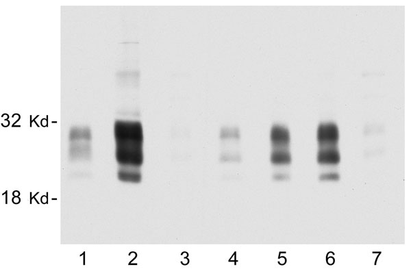
Figure 3. Representative immunoblot showing the relative amount of disease-associated prion protein (PrPres) in brain, tonsil, and selected lymph nodes from a single chronic wasting disease (CWD)–affected mule deer. All lanes were loaded with 10-mg equivalents of tissue (original wet weight basis). Lane 1, brain; lane 2, tonsil; lane 3, popliteal lymph node; lane 4, retropharyngeal lymph node (RPLN); lane 5, prescapular lymph node; lane 6, submandibular lymph node; lane 7, mesenteric lymph node. PrPres bands were visualized by using antibody L42 at 0.04 μg/mL and standard enhanced chemiluminescence processing.
Page created: June 28, 2010
Page updated: June 28, 2010
Page reviewed: June 28, 2010
The conclusions, findings, and opinions expressed by authors contributing to this journal do not necessarily reflect the official position of the U.S. Department of Health and Human Services, the Public Health Service, the Centers for Disease Control and Prevention, or the authors' affiliated institutions. Use of trade names is for identification only and does not imply endorsement by any of the groups named above.