Volume 13, Number 6—June 2007
Research
Levels of Abnormal Prion Protein in Deer and Elk with Chronic Wasting Disease
Brent L. Race*, Kimberly D. Meade-White*, Anne Ward*, Jean Jewell†, Michael W. Miller‡, Elizabeth S. Williams†1, Bruce Chesebro*, and Richard E. Race*
Figure 4
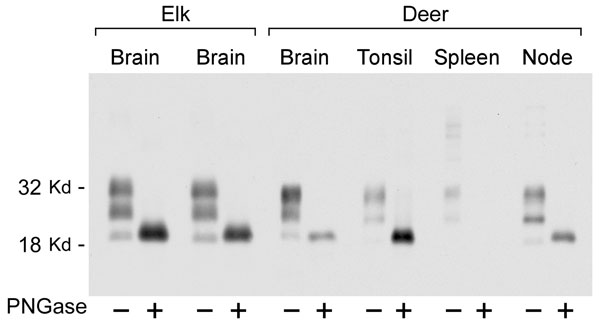
Figure 4. Immunoblot showing disease-associated prion protein from chronic wasting disease–affected elk brain or mule deer brain, tonsil, spleen, and retropharyngeal lymph node before and after treatment with PNGaseF. Alternating lanes show before and after treatment for each tissue. PNGaseF digestion was done as described in Materials and Methods. Ten-milligram equivalents of tissue were used for PNGase F–negative lanes, and 4-mg equivalents of tissue were used for PNGase F–positive lanes. The blot was developed as described in Figure 3.
Page created: June 28, 2010
Page updated: June 28, 2010
Page reviewed: June 28, 2010
The conclusions, findings, and opinions expressed by authors contributing to this journal do not necessarily reflect the official position of the U.S. Department of Health and Human Services, the Public Health Service, the Centers for Disease Control and Prevention, or the authors' affiliated institutions. Use of trade names is for identification only and does not imply endorsement by any of the groups named above.