Skip directly to site content Skip directly to page options Skip directly to A-Z link Skip directly to A-Z link Skip directly to A-Z link
Volume 14, Number 11—November 2008
Dispatch

Use of Malaria Rapid Diagnostic Test to Identify Plasmodium knowlesi Infection

Thomas F. McCutchanComments to Author , Robert C. Piper, and Michael T. Makler
Author affiliations: National Institute of Allergy and Infectious Diseases, Bethesda, Maryland, USA (T.F. McCutchan); University of Iowa, Iowa City, Iowa, USA (R.C. Piper); Flow Incorporated, Portland, Oregon, USA (M.T. Makler)

Main Article

Figure 2

Modeling of the analysis of Plasmodium knowlesi lactate dehydrogenase (LDH). A) Sequence of LDH from P. knowlesi deduced from genomic DNA fragments sequenced by the Sanger malaria genome project (www.sanger.ac.uk/Projects/P_knowlesi). LDH isoforms from P. vivax, P. malariae, P. ovale, P. berghei, P. yoelli, and P. falciparum were compared with that of P. knowlesi. Residues unique to P. knowlesi and P. vivax are shown in blue; residues unique to P. knowlesi and P. falciparum are shown in red. B)

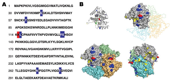
Figure 2. Modeling of the analysis of Plasmodium knowlesi lactate dehydrogenase (LDH). A) Sequence of LDH from P. knowlesi deduced from genomic DNA fragments sequenced by the Sanger malaria genome project (www.sanger.ac.uk/Projects/P_knowlesi). LDH isoforms from P. vivax, P. malariae, P. ovale, P. berghei, P. yoelli, and P. falciparum were compared with that of P. knowlesi. Residues unique to P. knowlesi and P. vivax are shown in blue; residues unique to P. knowlesi and P. falciparum are shown in red. B) Model of P. knowlesi LDH and specific epitopes. A model for P. knowlesi LDH was calculated by using WURST protein threading server (www.zbh.uni-hamburg.de/wurst/index.php) and the P. falciparum and P. vivax crystal structures (PDB: 2A94 and 3 2AA3). Shown is the monomer, as well as the assembled tetramer, aligned to the backbone of the P. vivax tetramer using pymol. The nicotinamide adenine dinucleotide cofactor analog 3-acetyl pyridine adenine dinucleotide is shown in black. Residues important for substrate binding and catalysis are shown in yellow. P. knowlesi residues shared only with P. vivax are shown in blue and indicate where the 11D9/13H11 epitopes could be. P. knowlesi residues shared only with P. falciparum are shown in red and indicate a critical determinant of the 17E4/7G9 epitopes.

Main Article

Page created: July 21, 2010
Page updated: July 21, 2010
Page reviewed: July 21, 2010
The conclusions, findings, and opinions expressed by authors contributing to this journal do not necessarily reflect the official position of the U.S. Department of Health and Human Services, the Public Health Service, the Centers for Disease Control and Prevention, or the authors' affiliated institutions. Use of trade names is for identification only and does not imply endorsement by any of the groups named above.
file_external