Volume 15, Number 9—September 2009
Research
Genetics and Pathogenesis of Feline Infectious Peritonitis Virus
Meredith A. Brown , Jennifer L. Troyer, Jill Pecon-Slattery, Melody E. Roelke, and Stephen J. O’Brien
Figure 2
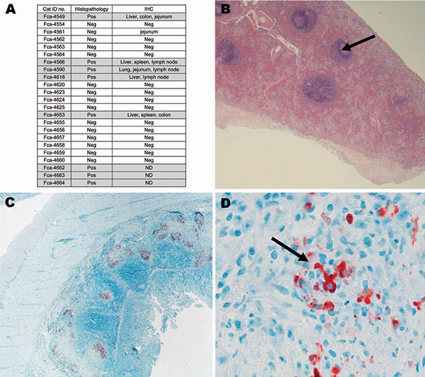
Figure 2. A) Histopathologic and immunohistochemical (IHC) results from 23 necropsied cats positive for antibodies against feline coronavirus. Liver, lung, spleen, colon, jejunum, stomach, heart, kidney, lymph node were evaluated by IHC. Feline infectious peritonitis (FIP) cases are highlighted in gray. Pos, positive; Neg, negative; ND, not done. B) Representative tissues from cat no. FCA-4653, spleen (histopathologic) showing granuloma (arrow); magnification ×20. C) Representative tissues from cat no. FCA-4590, small intestine (IHC); magnification ×20. D) Red staining indicates binding of coronavirus antibody (CoV p56, arrow); magnification ×100.
Page created: December 07, 2010
Page updated: December 07, 2010
Page reviewed: December 07, 2010
The conclusions, findings, and opinions expressed by authors contributing to this journal do not necessarily reflect the official position of the U.S. Department of Health and Human Services, the Public Health Service, the Centers for Disease Control and Prevention, or the authors' affiliated institutions. Use of trade names is for identification only and does not imply endorsement by any of the groups named above.