Volume 15, Number 9—September 2009
Dispatch
Rickettsia parkeri in Amblyomma americanum Ticks, Tennessee and Georgia, USA
Cite This Article
Citation for Media
Abstract
To determine the geographic distribution of the newly recognized human pathogen Rickettsia parkeri, we looked for this organism in ticks from Tennessee and Georgia, USA. Using PCR and sequence analysis, we identified R. parkeri in 2 Amblyomma americanum ticks. This rickettsiosis may be underdiagnosed in the eastern United States.
The most commonly reported rickettsial human pathogen in the United States is Rickettsia rickettsii, the causative agent of Rocky Mountain spotted fever (RMSF). First identified in 1937 (1), R. parkeri has been recognized as a human pathogen only since 2004, when it was isolated from an eschar on a serviceman from Virginia (2), although an Ohio patient suspected to have RMSF died of R. parkeri rickettsiosis in 1990 (3). Little is known about the geographic distribution of R. parkeri in the United States or the epidemiology of the disease it causes. The primary vector of R. parkeri is thought to be the Gulf Coast tick (Amblyomma maculatum), and naturally infected Gulf Coast ticks have been reported in numerous southeastern states (4). Experimentally, A. americanum ticks can maintain and transmit R. parkeri (5); thus, this tick species, which is more abundant than A. maculatum and bites humans aggressively (6), might contribute to R. parkeri transmission. To determine the geographic distribution of R. parkeri, we examined ticks collected in Tennessee and Georgia.
In Georgia, during 2005–2006, residents were encouraged to submit ticks to the state’s Division of Public Health for identification and testing for various tick-borne pathogens. We studied only ticks that had been attached to persons. Ticks were individually homogenized with metal beads and resuspended in 225 μL of phosphate-buffered saline. DNA was extracted from 100 μL of the homogenate by using a QIAamp DNA Micro Kit (QIAGEN Inc, Valencia, CA, USA) according to the manufacturer's instructions. We conducted a nested PCR targeting the 17-kDa gene (7) and used bidirectional sequencing at the Integrated Biotechnology Laboratories (University of Georgia, Athens, GA, USA) to confirm positive results.
In Tennessee, from April 2007 through September 2008, the United States Department of Agriculture, Animal and Plant Health Inspection Service, Wildlife Services, and the Tennessee Department of Health collected A. americanum and A. maculatum ticks from 31 counties by examining wild animals and dragging flannel sheets through vegetation. Ticks were stored in 100% ethanol and were sent to the Tennessee Department of Health Vector-Borne Diseases Laboratory for identification of species and life stage and detection of pathogens by molecular testing. A PCR targeting the outer membrane protein A (rOmpA) gene of spotted fever group (SGF) rickettsiae was conducted as previously described, using primers Rr190.602n and Rr190.70p (8). To identify the rickettsiae species, we subjected positive samples to a restriction fragment length polymorphism (RFLP) assay by digestion of the amplicons with RsaI (Promega, Madison, WI, USA) and PstI (Fermentas, Glen Burnie, MD, USA) enzymes at 37°C for 2 h. Digested fragments were subjected to electrophoresis on 10% polyacrylamide gels. To confirm species identification, we purified representative positive PCR products with Exosap-It (USB Corporation, Cleveland, OH, USA), sequenced at the Tennessee Department of Health Laboratory Services (Nashville, TN, USA) and entered into the National Center for Biotechnology Information BLAST database (www.blast.ncbi.nlm.nih.gov/Blast.cgi).
Of ticks collected in Georgia, 418 A. americanum ticks (237 adults, 180 nymphs, and 1 unknown) and 19 A. maculatum ticks, were submitted for testing. Of these, 1 A. americanum tick (Fayette County, May 2005) and 1 A. maculatum tick (Morgan County, July 2005) were positive for R. parkeri (100% identity with GenBank accession no. U17008).
Of 611 A. americanum and 2 A. maculatum ticks collected in Tennessee, 446 A. americanum (164 adults and 282 nymphs) and 2 A. maculatum adults were individually tested for Rickettsia spp. An additional 103 A. americanum larvae were divided in 10 pools of 4–19 ticks each according to collection site. A single A. americanum adult male (0.2% of total) had a positive RFLP pattern that matched the previously described rOmpA gene pattern of R. parkeri (8). The sequence of this amplicon (532 bp, GenBank accession no. FJ793521) was 99% similar to that of R. parkeri (EU715288). The positive tick had been collected from a coyote in Knox County, Tennessee, in July 2007.
Figure
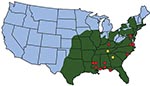
Figure. Location of ticks, Rickettsia parkeri in ticks, and human cases of rickettsiosis in the United States. Green shading indicates approximate distribution of Amblyomma americanum ticks, which completely overlaps with the known...
We identified R. parkeri in ticks in Tennessee and Georgia. In Tennessee, our identification of only 2 A. maculatum ticks (the primary vector of R. parkeri) supports previous reports that this tick species is uncommon in Tennessee (9). In Georgia, the 19 A. maculatum ticks identified were <5% of the total number of ticks collected in Georgia. In contrast to these low numbers, A. americanum ticks are ubiquitous in high densities throughout the other southeastern states and readily feed on humans (8). Recently, the range of A. americanum ticks has expanded in the United States and now extends from west-central Texas to the Atlantic Coast, encompassing the entire Southeast and parts of the lower Midwest and coastal New England (10). The distribution of A. americanum ticks completely overlaps the suspected distribution of A. maculatum ticks (Figure). The positive A. americanum tick from Tennessee was collected from a free-ranging coyote, and the detected R. parkeri may have been present in the blood meal taken by the tick. However, A. americanum ticks can maintain and transmit R. parkeri infection both transovarially and transtadially (6). R. parkeri has previously been identified in A. maculatum ticks from Georgia (4), but the identification of R. parkeri in A. americanum ticks renews concerns that this tick species may be involved in the natural history of another zoonotic pathogen. Additional study is needed to determine the extent of the role of A. americanum ticks as a natural vector for R. parkeri.
This study raises concerns about the serologic diagnosis of RMSF. R. parkeri may be the etiologic agent of some rickettsiosis cases in Tennessee and Georgia that have been misdiagnosed as RMSF. Because reliable clinical tests specific for different SFG rickettsiae are not readily available, several different rickettsioses may be serologically cross-reactive, leading to misdiagnosis of RMSF (11–14). Reliable diagnosis requires PCR or culture of biopsy specimens from eschars, when present (15). Additional studies characterizing SFG rickettsioses, including development of rickettsial species–specific clinical tests, will assist in attributing rickettsiosis to R. rickettsii, R. parkeri, or other SFG rickettsial infections.
Ms Cohen completed this work as part of an Emerging Infectious Diseases Laboratory Training Fellowship at the Tennessee Department of Health. She is entering a doctoral degree program at Cornell University to pursue her interests in infectious disease.
Acknowledgment
This research was supported by an appointment to the Emerging Infectious Diseases Fellowship Program administered by the Association of Public Health Laboratories and funded by the Centers for Disease Control and Prevention.
References
- Parker RR, Hohls GM, Cox GW, David GE. Observations on an infectious agent from Amblyomma maculatum. Public Health Rep. 1939;54:1482–4.
- Paddock CD, Sumner JW, Comer JA, Zaki SR, Goldsmith CS, Goddard J, Rickettsia parkeri: a newly recognized cause of spotted fever rickettsiosis in the United States. Clin Infect Dis. 2004;38:805–11. DOIPubMedGoogle Scholar
- Ralph D, Pretzman C, Daugherty N, Poetter K. Genetic relationships among the members of the family Rickettsiaceae as shown by DNA restriction fragment polymorphism analysis. Ann N Y Acad Sci. 1990;590:541–52. DOIPubMedGoogle Scholar
- Sumner JW, Durden LA, Goddard J, Stromdahl EY, Clark KL, Reeves WK, Gulf Coast ticks (Amblyomma maculatum) and Rickettsia parkeri, United States. Emerg Infect Dis. 2007;13:751–3.PubMedGoogle Scholar
- Goddard J. Experimental infection of lone star ticks, Amblyomma americanum (L.), with Rickettsia parkeri and exposure of guinea pigs to the agent. J Med Entomol. 2003;40:686–9.PubMedGoogle Scholar
- Merten HA, Durden LA. A state-by-state survey of ticks recorded from humans in the United States. J Vector Ecol. 2000;25:102–13.PubMedGoogle Scholar
- Labruna MB, Whitworth T, Bouyer DH, McBride JW, Camargo LMA, Camargo EP, Rickettsia bellii and Rickettsia amblyommii in Amblyomma ticks from the State of Rondonia, Western Amazon, Brazil. J Med Entomol. 2004;41:1073–81.PubMedGoogle Scholar
- Eremeeva M, Yu X, Raoult D. Differentiation among spotted fever group rickettsiae species by analysis of restriction fragment length polymorphism of PCR-amplified DNA. J Clin Microbiol. 1994;32:803–10.PubMedGoogle Scholar
- Durden LA, Kollars TM Jr. An annotated list of the ticks (Acari: Ixodoidea) of Tennessee, with records of four exotic species for the United States. Bulletin of the Society of Vector Ecology. 1992;17:125–31.
- Childs JE, Paddock CD. The ascendancy of Amblyomma americanum as a vector of pathogens affecting humans in the United States. Annu Rev Entomol. 2003;48:307–37. DOIPubMedGoogle Scholar
- Paddock CD, Finley RW, Wright CS, Robinson HN, Schrodt BJ, Lane CC, Rickettsia parkeri rickettsiosis and its clinical distinction from Rocky Mountain spotted fever. Clin Infect Dis. 2008;47:1188–96. DOIPubMedGoogle Scholar
- Apperson CS, Engber B, Nicholson WL, Mead DG, Engel J, Yabsley MJ, Tick-borne diseases in North Carolina: is “Rickettsia amblyommii” a possible cause of rickettsiosis reported as Rocky Mountain spotted fever? Vector Borne Zoonotic Dis. 2008;8:597–606. DOIPubMedGoogle Scholar
- Raoult D, Paddock CD. Rickettsia parkeri infection and other spotted fevers in the United States. N Engl J Med. 2005;353:626–7. DOIPubMedGoogle Scholar
- Parola P, Paddock CD, Raoult D. Tick-borne rickettsioses around the world: emerging diseases challenging old concepts. Clin Microbiol Rev. 2005;18:719–56. DOIPubMedGoogle Scholar
- Whitman TJ, Richards AL, Paddock CD, Tamminga CL, Sniezek PJ, Jiang J, Rickettsia parkeri infection after tick bite, Virginia. Emerg Infect Dis. 2007;13:334–6. DOIPubMedGoogle Scholar
Figure
Cite This ArticleTable of Contents – Volume 15, Number 9—September 2009
| EID Search Options |
|---|
|
|
|
|
|
|
Please use the form below to submit correspondence to the authors or contact them at the following address:
Abelardo C. Moncayo, Tennessee Department of Health, 630 Hart Ln, Nashville, TN 37216, USA;
Top