Volume 18, Number 11—November 2012
Dispatch
Vibrio fluvialis in Patients with Diarrhea, Kolkata, India
Cite This Article
Citation for Media
Abstract
We identified 131 strains of Vibrio fluvialis among 400 nonagglutinating Vibrio spp. isolated from patients with diarrhea in Kolkata, India. For 43 patients, V. fluvialis was the sole pathogen identified. Most strains harbored genes encoding hemolysin and metalloprotease; this finding may contribute to understanding of the pathogenicity of V. fluvialis.
Many members of the family Vibrionaceae cause diarrheal disease; among these, Vibrio cholerae O1/O139 and V. parahaemolyticus are responsible for several epidemics and pandemics (1,2). In Indonesia, >20% of diarrheal infections are caused by pathogenic Vibrio spp (3). Some of these Vibrio spp. can grow in thiosulfate–citrate–bile salts–sucrose agar as yellow colonies and do not agglutinate with V. cholerae O1 antiserum. These species are broadly defined as nonagglutinating (NAG) vibrios.
The emerging etiologic agent V. fluvialis has caused sporadic cases and outbreaks of diarrhea in several countries (4–6). Species-specific minimal biochemical tests, e.g., lysine decarboxylase, ornithine decarboxylase, arginine didydrolase, and L-arabinose, are used to identify V. fluvialis; without these tests, it may be confused with NAG vibrios, V. cholerae, and even Aeromonas spp. In most resource-poor countries, these tests are not performed, which may lead to labeling of V. fluvialis as a NAG vibrio.
Although V. fluvialis is known to cause diarrhea, the mechanisms involved in its pathogenicity are not well established. To evaluate the prevalence of V. fluvialis in India and possible mischaracterization as an NAG vibrio, we examined cases in which isolates from hospitalized patients with diarrhea were identified as NAG vibrios and characterized the strains using phenotypic and genetic methods.
We examined 400 isolates identified as NAG vibrios that were collected during 2002–2009 from 11,904 stool specimens from patients with diarrhea admitted to the Infectious Diseases and Beliaghata General Hospital, Kolkata, India. Specimens were screened for common enteric pathogens, according to standard protocols (7). Oxidase, string test, and arginine dihydrolase–positive strains that did not agglutinate with V. cholerae O1 polyvalent or O139 monovalent antiserum were further confirmed as V. fluvialis by using a multiplex PCR targeting the toxR gene of V. fluvialis and the ompW gene of V. cholerae (8,9). Isolates were also subjected to PCRs targeting different virulence-associated genes encoding the repeat in toxin (rtxA, rtxC), heat-stable enterotoxin (stn), type 3 secretion system (vcsC2, vcsV2, vcsN2 and vspD), cholera toxin (ctxA), toxin co-regulated pilus (tcpA), thermostable direct-hemolysin (tdh), TDH-related hemolysin (trh), V. fluvialis hemolysin (VFH), and metalloproteases, according to published methods (10–12).
Expression of VFH was determined in vitro by using erythrocytes from rabbit and sheep. Cytotoxin assay was performed with HeLa and Chinese hamster ovary cell lines by using sterile culture filters of the V. fluvialis strains that were isolated as a sole pathogen. Antimicrobial drug susceptibility testing was performed by using the disk diffusion method with commercially available disks (Becton Dickinson, Sparks Glencoe, MD, USA), according to Clinical and Laboratory Standards Institute criteria (13). Because these guidelines do not include interpretive criteria for V. fluvialis, breakpoints for Enterobacteriaceae were adopted. Escherichia coli ATCC 25922 was used as a quality control strain.
Pulsed-field gel electrophoresis was performed according to the PulseNet standardized protocol for V. cholerae (14). Gel Compare II software (Applied Maths NV, Sint-Martens-Latem, Belgium) was used for electrophoresis pattern comparison that runs on Dice similarity index and unweighted pairgroup with arithmetic mean method.
Among the 400 isolates presumptively identified NAG vibrios, multiplex PCR confirmed 131 and 269 strains (each strain representing a case) as V. fluvialis and V. cholerae, respectively. The overall prevalence rate of V. fluvialis among 11,904 hospitalized patients with diarrhea was 1.1%. Abrupt appearance of V. fluvialis was identified in 2002, although the surveillance of diarrheal infection was initiated at the Infectious Diseases and Beliaghata General Hospital in 1996 (www.niced.org.in/annual_reports.htm). The isolation rate of V. fluvialis gradually increased from 0.7% in 2002 to 2.2% in 2009 (Table 1). Of the 131 strains of V. fluvialis, 43 (33%) were identified as the sole pathogen; the remaining 88 (67%) were isolated as a co-pathogen with either V. cholerae, V. parahaemolyticus, E. coli, Shigella spp., parasites, or enteric viruses (data not shown). Among the mixed infections, V. fluvialis with V. cholerae was isolated most often (17%), followed by V. fluvialis and V. parahaemolyticus (8%). The presence of Vibrio spp. as mixed pathogens indicates that these patients likely acquired the infection from contaminated water or food. We analyzed the date of admission and place from where the patients resided and found no evidence for clusters of infection or small outbreaks caused by V. fluvialis.
V. fluvialis infection was much more often detected in adults (73%) than in children <5 years of age (27%). Clinical symptoms of sole infection caused by V. fluvialis were similar to that of cholera: watery diarrhea (86%), severe dehydration status (28%), and abdominal pain (12%) (Table 2). Several previous investigations have identified cholera-like diarrheal outbreaks caused by V. fluvialis (4,5).
All the V. fluvialis strains were negative for the virulence genes commonly reported in V. cholerae and V. parahaemolyticus, but >90% were positive for genes encoding VFH and metalloproteases. More than 80% of the strains expressed hemolysin against rabbit and sheep red blood cells. Hemolysin is a widely distributed virulence factor in most pathogenic Vibrio spp. Metalloprotease produced by V. fluvialis is related to hemagglutination proteases of V. vulnificus, which enhances permeability and hemorrhagic activities (12). These factors may increase the virulence of V. fluvialis and contribute to diarrhea.
When the culture filtrates were tested, cytotoxic effect was readily noticed in the Chinese hamster ovary and HeLa cell lines, i.e., cytoplasmic vacuolation, cell rounding, and destruction of the monolayer. In most strains isolated as a sole pathogen, the cytotoxic endpoint titer was 2–256 (Technical Appendix Table 1). The cell vacuolation phenomenon has been reported as a virulence factor in several enteric pathogens (Technical Appendix References).
In this study, V. fluvialis strains were highly resistant to ampicillin (92%), streptomycin (85%), furazolidone (85%), and sulfamethoxazole/trimethoprim (70%) (Technical Appendix Table 2). About half the number of strains were resistant to ciprofloxacin and 45% to nalidixic acid; the lower resistance rate for nalidixic acid compared with fluoroquinolones is unexpected and warrants further investigation to confirm the additional mechanisms. In a previous study, we found that some V. fluvialis strains carried the plasmid-mediated quinolone resistance gene allele qnrA1 and a gene encoding the aminoglycoside acetyltransferase (aac(6')-Ib-cr), which reduces ciprofloxacin activity (15). Fluroquinolone resistance and intermediate susceptibility to erythromycin (92%) are the unique features of the V. fluvialis isolated in this study; this trend was not recorded in other Vibrio spp., e.g., V. cholerae and V. parahaemolyticus.
Figure
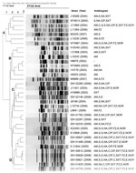
Figure. . . . Dendrogram of NotI-digested pulsed-field gel electrophoresis (PFGE) profiles with representative Vibrio fluvialis isolates. Clustering identified 4 clades (A–D). AM, ampicillin; S, streptomycin; G, gentamicin; NA, nalidixic acid; CIP,...
Although the V. fluvialis strains exhibited distinct NotI restriction profiles in the denrogroam analysis, at least 4 major clades were identified (Figure). Clades A and B, with strains isolated during 2002–2007, exhibited less antimicrobial drug resistance than did clade C and D strains identified during 2008–2009; multidrug-resistant strains, especially those resistant to fluroquinolones, were identified in higher numbers in clades C and D (Figure).
Our results demonstrate an emerging trend of prevalence of V. fluvialis among patients with acute diarrhea patients in Kolkata. The expression of cytotoxic activity and hemolysin may contribute to understanding the pathogenicity of V. fluvialis. Further epidemiologic studies are necessary to elucidate the public health importance of V. fluvialis–mediated diarrhea.
Mr Chowdhury is a doctoral candidate at the National Institute of Cholera and Enteric Diseases, Kolkata, India. His main research interest is the pathogenesis and molecular biology of enteric Vibrio spp.
Acknowledgment
This work was supported in part by the Ministry of Health, Labor and Family Welfare of Japan (Project H17-Shinkou-3); Initiative for Global Research Network on Infectious Diseases, Ministry of Education, Culture, Sports, Science and Technology, Japan; and intramural grants of the Indian Council of Medical Research, New Delhi, India.
References
- Nair GB, Ramamurthy T, Bhattacharya SK, Dutta B, Takeda Y, Sack DA. Global dissemination of Vibrio parahaemolyticus serotype O3:K6 and its serovariants. Clin Microbiol Rev. 2007;20:39–48. DOIPubMedGoogle Scholar
- Lesmana M, Subekti DS, Tjaniadi P, Simanjuntak CH, Punjabi NH, Campbell JR, Spectrum of Vibrio species associated with acute diarrhea in North Jakarta, Indonesia. Diagn Microbiol Infect Dis. 2002;43:91–7. DOIPubMedGoogle Scholar
- Huq MI, Alam AKMJ, Brenner DJ, Morris GK. Isolation of vibrio-like group EF-6, from patients with diarrhoea. J Clin Microbiol. 1980;11:621–4.PubMedGoogle Scholar
- Thekdi R, Lakhani AG, Vachha SM, Chandrakapure MR. Vibrio fluvialis (group F Vibrio) in Maharashtra. Indian J Med Res. 1982;76:80–5.PubMedGoogle Scholar
- Kobayashi K, Ohnaka T. Food poisoning due to newly recognized pathogens. Asian Med J. 1989;32:1–12.
- World Health Organization. Manual for laboratory identification of acute enteric infections. Geneva: The Organization; 1987.
- Chakraborty R, Sinha S, Mukhopadhyay AK, Asakura M, Yamasaki S, Bhattacharya SK, Species-specific identification of Vibrio fluvialis by PCR targeted to the conserved transcriptional activation and variable membrane tether regions of the toxR gene. J Med Microbiol. 2006;55:805–8. DOIPubMedGoogle Scholar
- Nandi B, Nandy RK, Mukhopadhyay S, Nair GB, Shimada T, Ghose AC. Rapid method for species-specific identification of Vibrio cholerae using primers targeted to the gene of outer membrane protein OmpW. J Clin Microbiol. 2000;38:4145–51.PubMedGoogle Scholar
- Chatterjee S, Ghosh K, Raychoudhuri A, Chowdhury G, Bhattacharya MK, Mukhopadhyay AK, Incidence, virulence factors, and clonality among clinical strains of non-O1, non-O139 Vibrio cholerae isolates from hospitalized diarrheal patients in Kolkata, India. J Clin Microbiol. 2009;47:1087–95. DOIPubMedGoogle Scholar
- Han JH, Lee JH, Choi YH, Park JH, Choi TJ, Kong IS. Purification, characterization and molecular cloning of Vibrio fluvialis hemolysin. Biochim Biophys Acta. 2002;1599:106–14. DOIPubMedGoogle Scholar
- Miyoshi S, Sonoda Y, Wakiyama H, Rahman MM, Tomochika K, Shinoda S, An exocellular thermolysin-like metalloprotease produced by Vibrio fluvialis: purification, characterization, and gene cloning. Microb Pathog. 2002;33:127–34. DOIPubMedGoogle Scholar
- Clinical and Laboratory Standards Institute. Performance standards for antimicrobial susceptibility testing. Document M100–S21. Wayne (PA): The Institute; 2011.
- Kam KM, Luey CK, Tsang YM, Law CP, Chu MY, Cheung TL, Molecular subtyping of Vibrio cholerae O1 and O139 by pulsed-field gel electrophoresis in Hong Kong: correlation with epidemiological events from 1994 to 2002. J Clin Microbiol. 2003;41:4502–11. DOIPubMedGoogle Scholar
- Chowdhury G, Pazhani GP, Nair GB, Ghosh A, Ramamurthy T. Transferable plasmid-mediated quinolone resistance in association with extended-spectrum β-lactamases and fluoroquinolone-acetylating aminoglycoside-6′-N-acetyltransferase in clinical isolates of Vibrio fluvialis. Int J Antimicrob Agents. 2011;38:169–73.PubMedGoogle Scholar
Figure
Tables
Cite This ArticleTable of Contents – Volume 18, Number 11—November 2012
| EID Search Options |
|---|
|
|
|
|
|
|
Please use the form below to submit correspondence to the authors or contact them at the following address:
Thandavarayan Ramamurthy, National Institute of Cholera and Enteric Diseases, P-33, CIT Road, Scheme XM, Beliaghata, Kolkata 700010, India
Top