Volume 18, Number 12—December 2012
Research
Virulent Avian Infectious Bronchitis Virus, People’s Republic of China
Figure 5
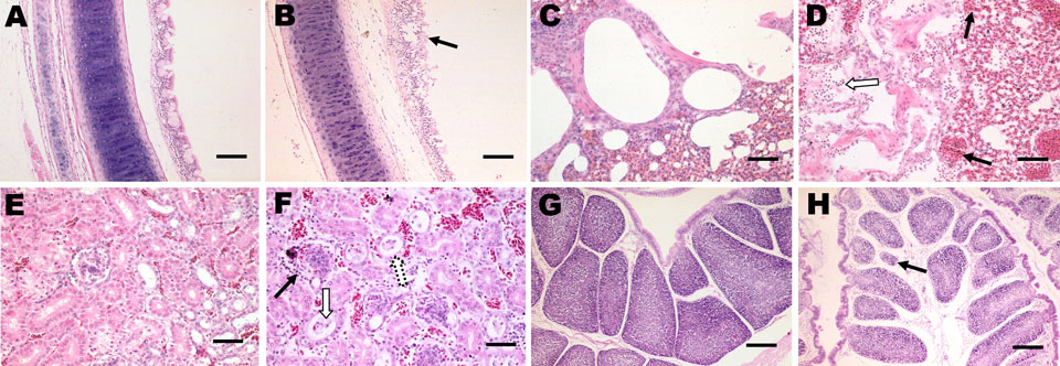
Figure 5. . Histopathologic analysis (hematoxylin and eosin stain) of tissues from 30-day-old chickens infected with infectious bronchitis virus YN strain. Panels A, C, E, and G, correspond to control tissues. B) Trachea, extensive dropout, degeneration, and necrosis of the ciliated epithelial cells (black arrow). Scale bar = 100 μm. D) Lung tissue with hemorrhage (black arrow), congestion, and lymphocytic infiltration in alveolar lumen (white arrow). Scale bar = 50 μm. F) Kidney tissue with severe renal lesions, including degeneration (white arrow), and necrosis of renal tubular epithelial cells, lymphocytic infiltration in the interstitium (black arrow), exfoliated renal tubular epithelial cells and erythrocytes were observed extensively. Scale bar = 50 μm. H) Bursa tissue with serous atrophy of lymphoid follicles and widening of the interstitium were observed in bursa of Fabricius (black arrow). Scale bar = 200 μm.