Volume 18, Number 12—December 2012
Research
Virulent Avian Infectious Bronchitis Virus, People’s Republic of China
Jinling Feng, Yanxin Hu, Zhijun Ma, Qi Yu, Jixun Zhao, Xiaodong Liu, and Guozhong Zhang
Figure 6
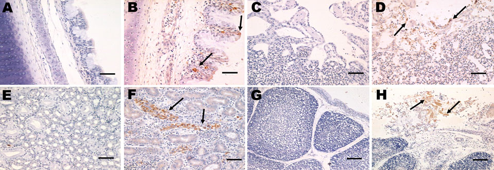
Figure 6. . Immunohistochemical detection of avian infectious bronchitis virus (IBV) antigens in tissues after experimental infection with IBV YN strain. Panels A, C, E, and G correspond to control tissues. B) Tracheal tissue with viral antigen detected extensively in the epithelial cells of the tracheal mucosa (black arrow). Scale bar = 50 μm. D) Lung tissue with viral antigen detected in alveolar cells (black arrow). Scale bar = 50 μm. F) Kidney tissue with viral antigens detected widely in the renal tubular epithelial cells (black arrow). Scale bar = 50 μm. H) Bursa tissue with viral antigens detected at high levels in mucosal epithelium in the bursa of Fabricius (black arrow). Scale bar = 100 μm.
Page created: November 21, 2012
Page updated: November 21, 2012
Page reviewed: November 21, 2012
The conclusions, findings, and opinions expressed by authors contributing to this journal do not necessarily reflect the official position of the U.S. Department of Health and Human Services, the Public Health Service, the Centers for Disease Control and Prevention, or the authors' affiliated institutions. Use of trade names is for identification only and does not imply endorsement by any of the groups named above.