Volume 19, Number 2—February 2013
CME ACTIVITY - Synopsis
Eastern Equine Encephalitis in Children, Massachusetts and New Hampshire,USA, 1970–2010
Figure 1
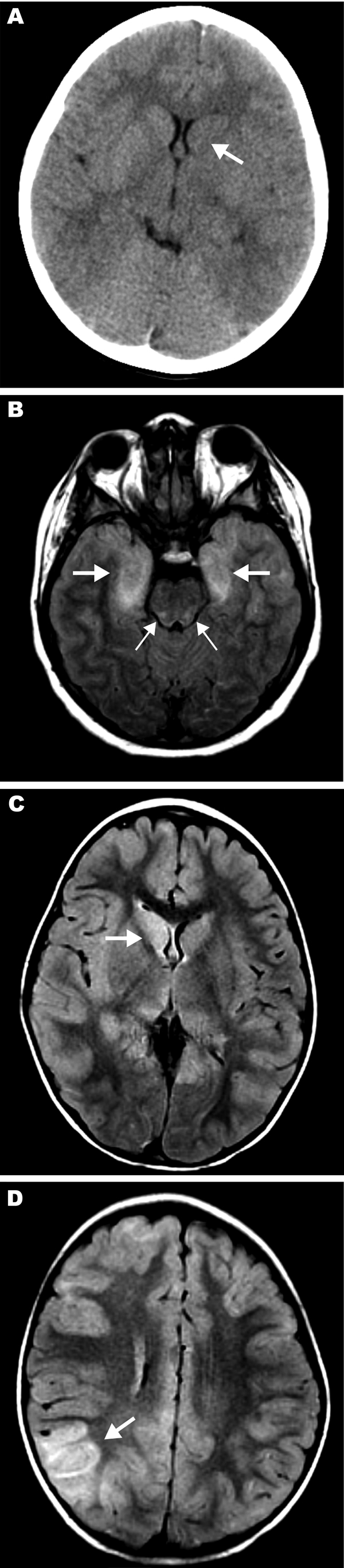
Figure 1. . . . . . Magnetic resonance images (MRIs) and computed tomography neuroradiographs showing lesions in brains of 3 children with eastern equine encephalitis A) Results of noncontrast computed tomography scan of the brain of patient 12 on hospital day 2; the neuroradiograph shows subtle hypoattenuation of the left caudate head (arrow) and diencephalic region. B) Axial fluid attenuated inversion recovery (FLAIR) image from brain MRI scan on patient 14 on hospital day 2; the image shows abnormal T2 hyperintense regions of the bimesial temporal regions (thick arrows) with accompanying abnormal T2 hyperintense regions of the dorsal pontomesencephalic regions (thin arrows). C, D) FLAIR images from brain MRI scan of patient 15 on hospital day 3. C) Abnormal T2 hyperintense caudate and thalamic nuclei, most prominent on the right (arrow). D) Abnormal T2 hyperintense regions are most prominent in the right parietotemporal gray matter (arrow) and subcortical white matter but are also seen scattered throughout.