Volume 19, Number 2—February 2013
CME ACTIVITY - Synopsis
Eastern Equine Encephalitis in Children, Massachusetts and New Hampshire,USA, 1970–2010
Figure 4

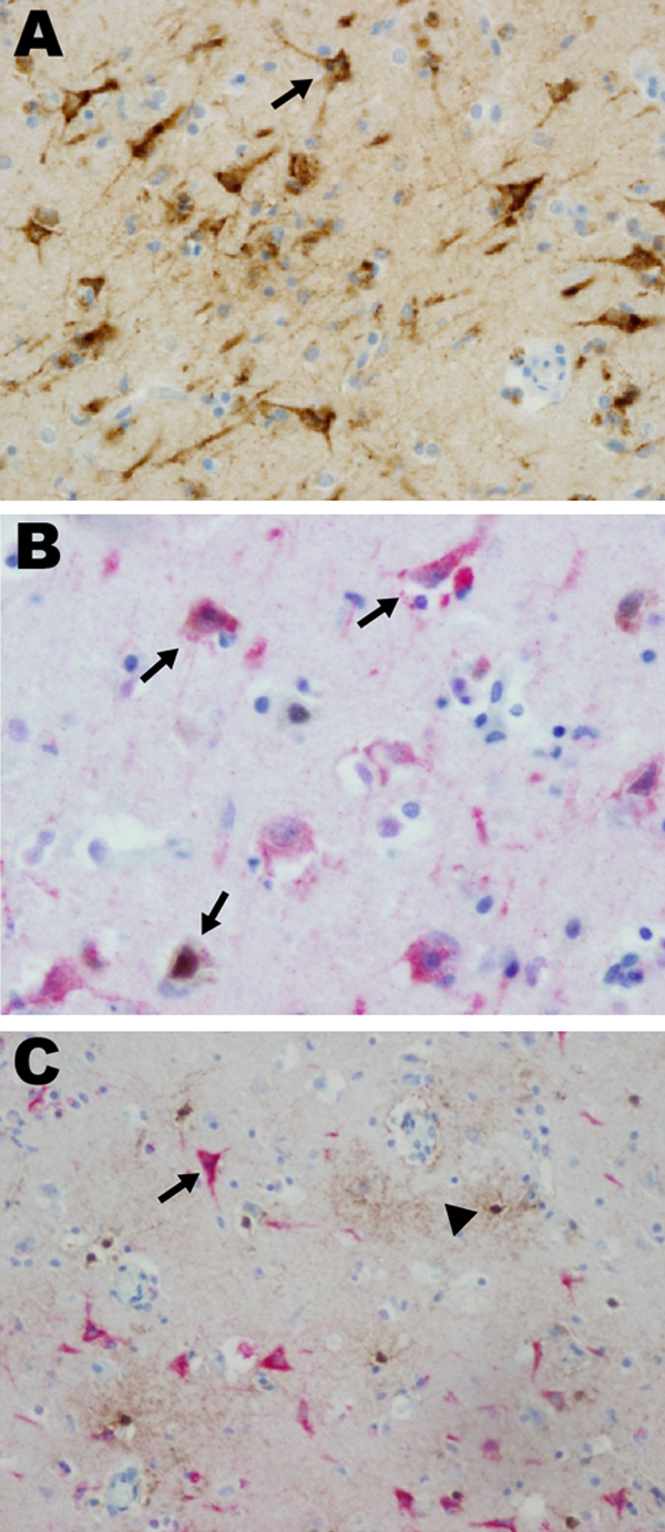
Figure 4. . . . . . Eastern equine encephalitis virus (EEEV) colocalized with neurons in patient 12 in a study of children with eastern equine encephalitis (EEE), Massachusetts and New Hampshire, 1970–2010. A) Immunohistochemistry, using EEEV immune ascites, of the entorhinal temporal cortex, demonstrating EEEV-infected neurons (arrow) (magnification ×400). B) Dual immunohistochemistry with EEE immune ascites (red stain) and a mouse monoclonal anti–neuronal nuclei (NeuN) antibody (brown stain) demonstrates that EEE-infected cells are NeuN–expressing neurons (arrows) (magnification ×400). C) Dual immunohistochemistry with EEE immune ascites (red stain) and rabbit polyclonal anti-glial fibrillary acidic protein (GFAP) antibody (brown stain) demonstrated that EEE-infected cells (arrow) do not express GFAP and are therefore not glial cells (arrowhead) (magnification ×400). Single antibody immunohistochemistry was performed with heat-induced antigen retrieval, using pressure cooker treatment (120°C for 30 s at 15 psi) with citrate buffer, pH 6.0. Lyophilized ascites from American Type Culture Collection (Manassas, VA, USA) was resuspended in 1 mL of double-distilled H20 and stored as stock solutions at −20°C. The ascites stocks were applied at 1:500 for 40 min at room temperature, after which labeled horseradish peroxidase (HRP) anti-mouse antibody was added, and the stocks were incubated 30 min at room temperature. Visualization was performed by using 3,3′-diaminobenzidine chromogen (Dako EnVision+ System-HRP (DAB); Dakocytomation, Carpinteria, CA, USA). Dual immunohistochemistry was performed by using polyclonal rabbit antibody to GFAP (DAKO, Carpinteria CA, USA) at 1:20,000 and mouse monoclonal anti-NeuN, clone A60, MAB377 (Millipore, Billerica, MA, USA) at 1:7,500. Both antibodies were visualized by using the Dako EnVision+ System-HRP (DAB)-brown, and then EEE immune ascites (1:500) was applied, using the alkaline phosphatase method, and visualized again by using Permanent Red (Dako).