Skip directly to site content Skip directly to page options Skip directly to A-Z link Skip directly to A-Z link Skip directly to A-Z link
Volume 19, Number 2—February 2013
CME ACTIVITY - Synopsis

Eastern Equine Encephalitis in Children, Massachusetts and New Hampshire,USA, 1970–2010

Michael A. Silverman, John Misasi, Sandra Smole, Henry A. Feldman, Adam B. Cohen, Sandro Santagata, Michael McManus, and Asim A. AhmedComments to Author 
Author affiliations: Author affiliations: Children's Hospital Boston, Boston, Massachusetts, USA (M.A. Silverman, J. Misasi, H.A. Feldman, S. Santagata, M. McManus, A.A. Ahmed); Department of Public Health, Boston (S. Smole); Massachusetts General Hospital, Boston (A.B. Cohen); Brigham and Women’s Hospital, Boston (S. Santagata)

Main Article

Figure 3

Histopathologic features for patient 12 in a study of children with eastern equine encephalitis (EEE), Massachusetts and New Hampshire, 1970–2010. The postmortem samples of central nervous system tissue were obtained 10 days after the onset of symptoms. A) Hematoxylin and eosin (H&E)–stained section of temporal lobe, showing meningeal inflammation (arrow) (magnification ×200). B) H&E-stained section of midbrain, showing perivascular inflammation (arrow) (magnification ×400). C–F) EEE vir

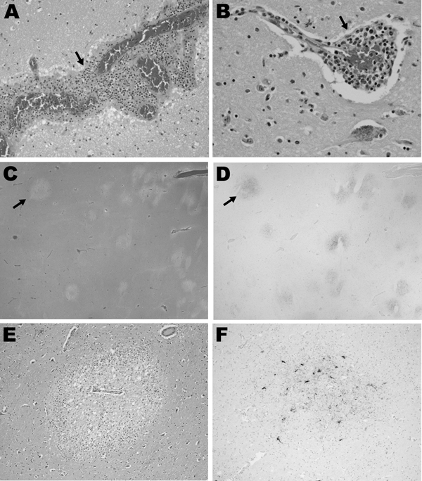
Figure 3. . . . . . Histopathologic features for patient 12 in a study of children with eastern equine encephalitis (EEE), Massachusetts and New Hampshire, 1970–2010. The postmortem samples of central nervous system tissue were obtained 10 days after the onset of symptoms. A) Hematoxylin and eosin (H&E)–stained section of temporal lobe, showing meningeal inflammation (arrow) (magnification ×200). B) H&E-stained section of midbrain, showing perivascular inflammation (arrow) (magnification ×400). C–F) EEE virus (EEEV) colocalizes with areas of tissue injury in the brain. C) H&E-stained section of the basal ganglia, demonstrating foci of marked tissue rarefaction (arrow) (magnification ×12.5). D) Immunohistochemistry of section adjacent to that shown in panel C; staining of the basal ganglia with EEE immune ascites demonstrates foci of EEEV (arrow) that correspond with areas of tissue rarefaction in panel C (magnification ×12.5). E) Immunohistochemistry with EEE immune ascites demonstrates EEE viral antigens in the thalamus. Specificity for EEEV immunoreactivity of this ascites fluid was confirmed by the lack of staining on control brain specimens (data not shown).

Main Article

Page created: January 17, 2013
Page updated: January 17, 2013
Page reviewed: January 17, 2013
The conclusions, findings, and opinions expressed by authors contributing to this journal do not necessarily reflect the official position of the U.S. Department of Health and Human Services, the Public Health Service, the Centers for Disease Control and Prevention, or the authors' affiliated institutions. Use of trade names is for identification only and does not imply endorsement by any of the groups named above.
file_external