Volume 19, Number 6—June 2013
Dispatch
Novel Poxvirus in Big Brown Bats, Northwestern United States
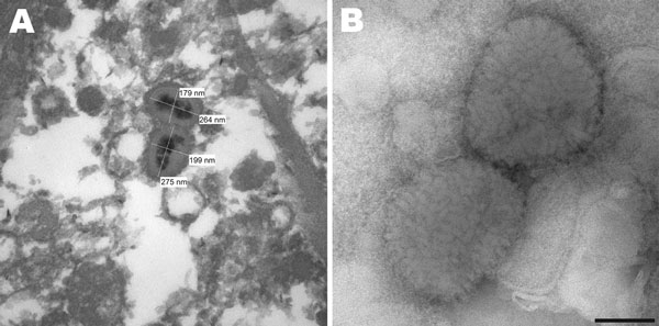
Figure 1

Figure 1. . . A) Electron micrograph of poxvirus particles in synovium of a big brown bat, northwestern United States. B) Negative staining of poxvirus particles in cell culture supernatant. Scale bar = 100 nm.
Page created: April 24, 2013
Page updated: April 24, 2013
Page reviewed: April 24, 2013
The conclusions, findings, and opinions expressed by authors contributing to this journal do not necessarily reflect the official position of the U.S. Department of Health and Human Services, the Public Health Service, the Centers for Disease Control and Prevention, or the authors' affiliated institutions. Use of trade names is for identification only and does not imply endorsement by any of the groups named above.